
Translate this page into:
Effect of bone bank processing on bone mineral density, histomorphometry & biomechanical strength of retrieved femoral head
Reprint requests: Dr O.P. Lakhwani, Department of Orthopaedics, Employees State Insurance Corporation - PostGraduate Institute of Medical Sciences and Research (ESI-PGIMSR), Basaidarapur, New Delhi 110 015, India e-mail: orth365@gmail.com
-
Received: ,
This is an open access journal, and articles are distributed under the terms of the Creative Commons Attribution-NonCommercial-ShareAlike 4.0 License, which allows others to remix, tweak, and build upon the work non-commercially, as long as appropriate credit is given and the new creations are licensed under the identical terms.
This article was originally published by Medknow Publications & Media Pvt Ltd and was migrated to Scientific Scholar after the change of Publisher.
Abstract
Background & objectives:
Standard processing of the bone grafts involves deep-freezing and sterilization with gamma irradiation which may alter mechanical properties of the bone graft. This study was aimed at measuring the effect of bone bank processing on the mechanical properties of bone allograft and its correlation with bone mineral density [BMD, dual-energy X-ray absorptiometry (DEXA Scan)] and histomorphometric indices.
Methods:
Femoral heads retrieved from patients undergoing hip replacement surgeries were used as the material. Twenty femoral heads were under taken in the study. Each femoral head was cut into two equal cubes. One cube was subjected to BMD measurement using DEXA Scan followed by unilateral compression test. Histomorphometric indices such as trabecular number (Tb. N.), trabecular separation (Tb. S.), trabecular thickness (Tb. T.) and bone volume (B.V.) were calculated on the same specimen by a computer software. The other cube was kept in deep freezer (−76°C) for a minimum of three weeks, followed by gamma irradiation and subjected to similar tests.
Results:
Results were compared in pre- and post-processed bone specimens. A significant loss of biomechanical strength (P<0.001) with mean a loss of 18.90 per cent was found in post-processed samples in uniaxial compression tests. Similarly, BMD (mean decrease by 13.8%, P<0.01) and histomorphometric indices such as Tb. T. (mean decrease by 12.37%, P<0.01), Tb. S. (mean increase by 12.60%, P<0.001) and B.V. (mean decrease by 20.84%, P<0.01) were found. However, Tb. N. was not significantly affected.
Interpretation & conclusions:
The current method of processing of bone allografts i.e. deep-freezing and gamma irradiation appeared to cause a significant reduction in the biomechanical strength of allogenic bone which was more suitable to be use in the morselized form. Appropriate consideration for decreased strength needs to be given when using allogenic bone graft as a structural graft.
Keywords
Biomechanical strength of bone
bone bank processing
bone mineral density
histomorphometry

Limited availability of the autologous bone graft and the high cost, non-biodegradability and lack of osteointegration attributed to synthetic bone graft substitutes have favoured the use of allogenic bone graft in osteo-reconstructive surgeries. Allogenic bone graft offers an attractive alternative to bone autograft as these are available in sufficient quantity without any donor-site morbidity12. Standard bone bank processing methods of allografts involve the use of deep-freezing and sterilization using gamma irradiation. Such processing methods may have an effect on the microstructural properties of allograft, which can be assessed by means of histomorphometry. So far, no study has been carried out to assess this effect by in vitro analysis.
Material engineering has demonstrated that all the material properties including mechanical properties are a function of material microstructure, in case of bone these being the trabeculae. Measures of apparent density [bone mineral density (BMD)] used as an estimate of mechanical properties does not account for the structural organization of the trabecular bone, thus rendering this scalar measure inadequate as a predictor of bone strength3. Studies have shown the insufficiency of BMD as a predictor of bone strength45. Thus, the evaluation of bone microstructure plays an important role in defining material properties of bone including the bone strength.
In vivo studies carried by Kang and Kim6 utilized mechanical properties of transplanted bone allograft subjected to deep-freezing at different durations and found that the transplanted bone subjected to deep-freezing was significantly weaker compared to their mechanical controls, but these studies only used biomechanical compression strength as a single parameter representing bone strength and effect on microstructure was not assessed. This in vitro study was undertaken using the retrieved femoral heads from living donors processed in the bone bank (deep-freezing and gamma irradiation) as a substrate to study the effect of bone bank processing on BMD [measured by dual energy X-ray absorptiometry (DEXA)], histomorphometric characteristics and compressive mechanical bone strength.
Material & Methods
The study was conducted during 2012-2014 in the departments of Orthopedics, Pathology and Radiology at Employees State Insurance Hospital, Postgraduate Institute of Medical Sciences and Research, Basaidarapur, New Delhi, in association with the department of Mechanical Engineering, Delhi Technological University, New Delhi, India. Twenty femoral heads retrieved during surgical hip replacement procedures were made devoid of all soft tissue and cartilage and cut into two equal cubical pieces using the bone saw. Exclusion criteria included femoral head involved in infection, Paget's disease, rheumatoid arthritis, osteoarthritis, neoplastic conditions, pathological fractures and other pathological conditions, which were non-representative of the general population. The study was approved by the ethics committee of the institute.
Of the two equal pieces, one was subjected to DEXA Scan followed by unilateral compression test and finally histomorphometry. The other piece was kept for deep-freezing at −76°C for a minimum period of three weeks followed by gamma irradiation of 25 Gy and then subjected to similar tests. Areal BMD was measured for both pre- and post-processed bone sample using the DEXA scan and analyzed. Areal BMD was measured for both pre- and post-processed bone sample using the DEXA scan and analyzed.
The first piece of the bone sample was tested for the failure in compression tests using Universal Testing Machine (Model 4482), Instron Corporation, United States, at a constant unilateral deformation rate 0.5 mm/sec. The resultant-deformation curve was analyzed and yield point calculated. The test was also performed on the bone bank processed piece of the cancellous bone sample after bringing it to the room temperature in a bath of normal saline using a similar procedure.
Bone histomorphometry is a quantitative histological examination of bone to access microstructure and predicts the biomechanical strength. Both pre- and post-processed bone sample underwent the histomorphometric analysis.
Procedure: The cancellous bone graft material was decalcified using five per cent nitric acid for 10 days (solution changed every two days), dehydrated in ascending grades of alcohol, cleared in xylene and then embedded in paraffin. Paraffin sections of 5 μm were cut using microtome and stained with haematoxylin & eosin. The image was processed using NIKON-based computerized software (Nikon, Japan) (×40 magnification) and histomorphometric parameters were calculated.
Parameters undertaken to analyze the structure (static parameters) were as follows:
(i) Bone volume (B.V.) (trabecular B.V./tissue volume) - represents the area occupied by the trabeculae vs the total area of the field. Trabecular B.V. (volume of total cancellous bone) was measured and expressed as a percentage.
(ii) Trabecular number (Tb. N.) - the number of trabeculae per unit area. In this study, single, high-power field at ×40 magnification was taken as a unit area.
(iii) Trabecular thickness (Tb. T.) - mean Tb. T. was measured and analyzed.
(iv) Trabecular separation (Tb. S.) - mean of the average distance between the two trabeculae was measured and analyzed.
The measured values of these indices in the given sample population are provided in the Table.

Statistical analysis: Paired t test was used to compare the pre- and post-processed samples using SPSS software, Version 19 (IBM Corp., USA).
Results
On comparing the BMD measurements between the pre-processed and the processed samples, a significant (P<0.01) decrease in BMD values as measured by DEXA was found. The mean decrease in BMD to the tune of 13.8 per cent was found. The effect of bone bank processing on microstructure was estimated using standard microscopy at ×40 magnification using standardized computerized software and the following parameters were studied: Tb.N., Tb. T., Tb.S. and B.V. (Table). Tb. N. i.e. number of trabeculae seen on a single ×40 field was measured in both pre- and post-processed (deep-freezing followed by gamma irradiation) samples. The processing of bone allograft did not cause a significant alteration in the microstructural parameter measured (Tb. N.). The Tb. T. was measured in both pre- and post-processed samples. The effect of bone bank processing on Tb. T. was measured and processing of bone allograft caused a significant (P<0.01) negative effect on the microstructural parameter measured. The mean decrease in Tb. T. was about 12.37 per cent.
The effect of bone bank processing on Tb.S. was measured and processing of bone allograft caused a significant (P<0.001) positive effect on the microstructural parameter measured. The mean increase in Tb. S. was found to be about 12.60 per cent. B.V. was measured and expressed as percentage in both pre- and post-processed samples. The processing of bone allograft showed a significant (P<0.01) negative effect on the microstructural parameter of measured B.V. The mean decrease in B.V. was found to be about 20.84 per cent. The Figure gives the actual view and measurements done at ×40 magnification.
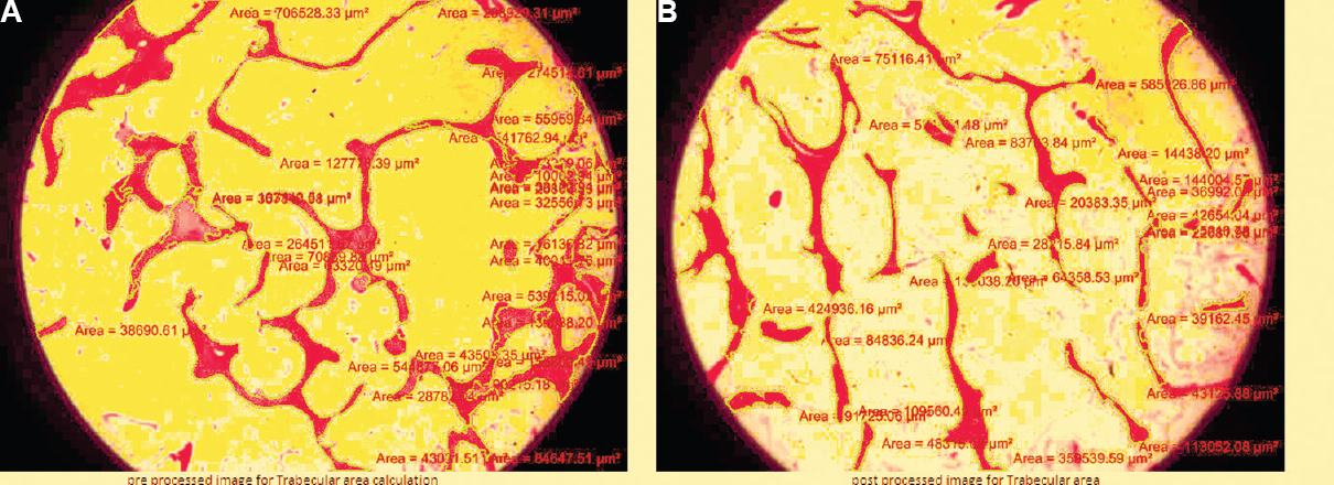
- Comparative histomorphometric pictures used for calculating Trabecular Area / Bone Volume of the pre- and post-processed samples showing decreased Trabecular Area / Bone Volume in post-processed image. (A) Pre-processed image (18.24%) and (B) post-processed image (13.32%).
Equal cubes obtained from the retrieved femoral head were tested for failure in compression tests, and the deformation curve was analyzed and yield point calculated in both pre- and post-processed samples. The processing of bone allograft caused a significant (P<0.001) negative effect on the compression strength of cancellous bone. The mean decrease in yield point was found to be about 18.90 per cent.
Discussion
It is postulated that the storage of allografts at low temperatures may affect the architecture of bone by producing microcracking, which may lead to increase in Tb. N. but at the same time causing loss of trabecular connectivity (decrease in Tb. T. and B.V. and increase in Tb. S.). Furthermore, radiation has an adverse effect on mechanical properties by damaging the collagen moiety of bone which may cause a loss of trabecular framework (decrease in Tb. N., Tb. T. and B.V. and increase in Tb. S.). Xu et al7 conducted an experimental study on rats and found a similar effect of radiation on microstructural parameters of bone. Similar results have been obtained in an experimental study using murine model8.
However, it has been postulated that deep-freezing of bone allograft only does not cause any appreciable change in densitometric and microstructural properties of bone allografts9. While BMD measurements may not get affected by deep-freezing alone9, high-dose gamma irradiation may have a negative effect on the same and the effect is cumulative when used with deep-freezing as a method to sterilize bone allografts. A significant decrease in BMD was found in post-processed samples as compared to pre-processed ones in our study.
Both deep-freezing and gamma irradiation caused a detrimental effect on Tb. T., and a loss of trabecular connectivity and hence an increase in Tb. S. Theoretically, deep-freezing may affect Tb. N. by producing microcracks while gamma irradiation may cause their disappearance. However, in this study, no significant difference in Tb. N. was observed between pre- and post-processed samples.
Several investigators studied mechanical properties of transplanted bone allograft subjected to deep-freezing at different durations and found that the transplanted bone subjected to deep-freezing was significantly weaker compared to their mechanical controls and that the effect on their mechanical properties was independent of the duration of deep freezing61011. A study by Kang and Kim6 using rat vertebrate model demonstrated a 20-23 per cent decrease in compressive strength following deep-freezing and the effect of deep freezing was independent of its duration.
Other studies have established the dose-dependent effect of gamma irradiation12131415. While standard dose that is 25 Gy did not have a significant effect on mechanical properties of bone allograft, higher dosages had significant detrimental effects on mechanical strength markers of bone. In the present study, the bone allografts were subjected to a minimum of three weeks duration of deep freezing at −76°C followed by gamma irradiation at 25 Gy and the results of processed sample to the pre-processed ones were compared. We found a significant decrease of 18.90 per cent in compressive strength of post-processed samples as compared to their controls. This result affirmed the detrimental effects of bone bank processing on the final bone strength.
Our study had some limitations. The sample size of the study was small and included patients undergoing hip replacement procedures which were non-representative of the entire population. A larger study incorporating larger sample size would be more conclusive so as to affirm and validate the results of this study.
In conclusion, our findings demonstrated that the current method of processing of bone allografts i.e. deep-freezing at −76°C for a minimum duration of three weeks followed by gamma irradiation at 25 Gy caused a significant reduction in biomechanical strength of allobone to the tune of 18.90 per cent. Thus, this method of processing is suitable in cases where the graft is intended to be used in the morselized form, but in cases wherein grafts are to be used as a structural graft, appropriate consideration needs to be given for the decrease in biomechanical strength. Newer chemical sterilization methods using peracetic acid ethanol16 have been proposed to retain the biomechanical properties of bone and act as an effective sterilization method and can be used in conditions where the graft is intended to be used as a structural graft.
Conflicts of Interest: None.
References
- Iliac crest bone graft harvest donor site morbidity. A statistical evaluation. Spine (Phila Pa 1976). 1995;20:1055-60.
- [Google Scholar]
- Donor site pain from the ilium. A complication of lumbar spine fusion. J Bone Joint Surg Br. 1989;71:677-80.
- [Google Scholar]
- The mechanical properties of trabecular bone: Dependence on anatomic location and function. J Biomech. 1987;20:1055-61.
- [Google Scholar]
- Hip fracture in women without osteoporosis. J Clin Endocrinol Metab. 2005;90:2787-93.
- [Google Scholar]
- Relationship between bone mineral density and bone stiffness in the bone fracture. Oral Radiol. 2005;21:38-40.
- [Google Scholar]
- The biomechanical properties of deep freezing and freeze drying bones and their biomechanical changes after in-vivo allograft. Yonsei Med J. 1995;36:332-5.
- [Google Scholar]
- The combined effects of X-ray radiation and hindlimb suspension on bone loss. J Radiat Res. 2014;55:720-5.
- [Google Scholar]
- A murine model for bone loss from therapeutic and space-relevant sources of radiation. J Appl Physiol. 2006;101:789-93.
- [Google Scholar]
- The mode of bone conservation does not affect the architecture and the tensile properties of rat femurs. Biomed Mater Eng. 2006;16:253-9.
- [Google Scholar]
- Biomechanical strength of deep-frozen versus lyophilized large cortical allografts. Clin Biomech (Bristol, Avon). 2004;19:526-33.
- [Google Scholar]
- Biomechanical strength of large diaphyseal deep-frozen allografts. Cell Tissue Bank. 2000;1:201-6.
- [Google Scholar]
- A comprehensive study of physical parameters, biomechanical properties, and statistical correlations of iliac crest bone wedges used in spinal fusion surgery. IV. Effect of gamma irradiation on mechanical and material properties. Spine (Phila Pa 1976). 1994;19:304-8.
- [Google Scholar]
- Biochemical properties of cortical allograft bone using a new method of bone strength measurement. A comparison of fresh, fresh-frozen and irradiated bone. J Bone Joint Surg Br. 1996;78:363-8.
- [Google Scholar]
- In vivo biomechanical effects of gamma irradiation on fresh-frozen allografts. Orthopedics. 1996;19:649-53.
- [Google Scholar]
- Effect of gamma irradiation on mechanical properties of human cortical bone: Influence of different processing methods. Cell Tissue Bank. 2012;13:363-74.
- [Google Scholar]
- Comparative biomechanical and microstructural analysis of native versus peracetic acid-ethanol treated cancellous bone graft. Biomed Res Int. 2014;2014:784702.
- [Google Scholar]


