
Translate this page into:
Paraspinal mass in thalassaemia is often extramedullary haematopoiesis
*For correspondence: hiralal2007@yahoo.co.in
This is an open access article distributed under the terms of the Creative Commons Attribution NonCommercial ShareAlike 3.0 License, which allows others to remix, tweak, and build upon the work non commercially, as long as the author is credited and the new creations are licensed under the identical terms.
This article was originally published by Medknow Publications & Media Pvt Ltd and was migrated to Scientific Scholar after the change of Publisher.
A 57 year old male, ex-smoker male who was a diagnosed case of beta-thalassaemia intermedia since last 22 years, presented to the department of Pulmonary Medicine, Sanjay Gandhi Post Graduate Institute of Medical Sciences, Lucknow, India, in January 2014. He had breathlessness on exertion and cough for the last two months. There was no past history of tuberculosis. The patient was on supportive care only for beta-thalassaemia with no requirement of blood transfusion. X-ray chest showed bilateral mediastinal masses (Fig. 1). Further, on non-contrast computed tomography (CT) and magnetic resonance imaging (MRI), these masses showed fat within the paraspinal mass (Figs. 2#x2013;4) which confirmed extramedullary haematopoiesis (EMH). The patient was symptomatically improved with supportive care and was called for follow up after three months, but was lost to follow up. Thoracic paraspinal masses, hepatosplenomegaly or focal lesions in liver, spleen and renal pelvis signify EMH in such patients. Knowledge of this entity is important to avoid misdiagnosing these as tumours.
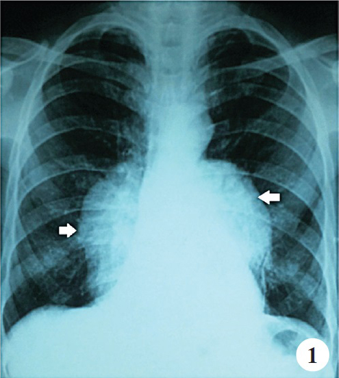
- Chest X-ray posterior anterior (PA) view shows bilateral well defined smooth convex outline radio-opacity seen in lower thoracic paraspinal area extending below the margin of diaphragm (white arrows)
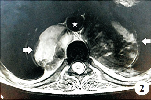
- MRI scan T2 weighted of thorax showing bilateral paraspinal mass lesions without intraspinal extension (white arrows). Aorta (marked by asterisk) is lifted away from the spine by the mass
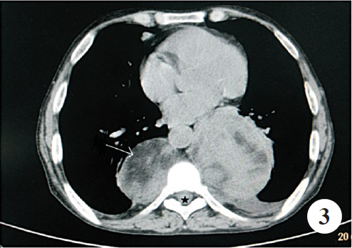
- Axial non-contrast computed tomography of the chest demonstrates fat withing the parasinal mass (marked by long arrow)
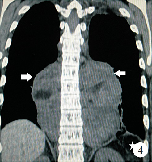
- Coronal non-contrast computed tomography of the chest demonstrates paraspinal mass on either side (marked as white arrow). Spleen is not visualized in splenic fossa (marked as asterisk). Patient had undergone splenectomy earlier.


