
Translate this page into:
Multiple papillomas in oral cavity of a six year old child
* For correspondence: drsjayachandranmds@yahoo.com
-
Received: ,
This is an open access journal, and articles are distributed under the terms of the Creative Commons Attribution-NonCommercial-ShareAlike 4.0 License, which allows others to remix, tweak, and build upon the work non-commercially, as long as appropriate credit is given and the new creations are licensed under the identical terms.
This article was originally published by Wolters Kluwer - Medknow and was migrated to Scientific Scholar after the change of Publisher.
A six year old boy† was brought to the department of Oral Medicine and Radiology of the Tamil Nadu Government Dental College, Chennai, India, in December 2017 with a complaint of growths on his upper and lower front teeth region for the past one month. On clinical examination, there were two pinkish pedunculated growths: 1 on the incisive papilla and the other on the attached gingiva of erupting mandibular central incisors (Fig. 1). A similar growth was earlier noticed on his tongue tip which got severed while he was playing. Within a period of one week thereafter, a similar growth was observed on the incisive papilla region which grew in size gradually in the next two weeks and then started contacting attached gingiva of erupting mandibular central incisors. After one week, another growth arising from the attached gingiva of erupting mandibular central incisors was noticed. No similar lesions were present elsewhere on the patient's body, and the patient's medical history was non-contributory. The growths were clinically diagnosed as papillomas and excised surgically. The histopathological report confirmed the clinical diagnosis of squamous papilloma (Figs 2 and 3). Usually, the oral papillomas present as single entity with very less virulence and no contagious potential. In this case there were multiple sites of occurrence including tongue, incisive papilla and lower mandibular attached gingiva. Three month follow up showed satisfactory healing and no recurrence (Fig. 4).
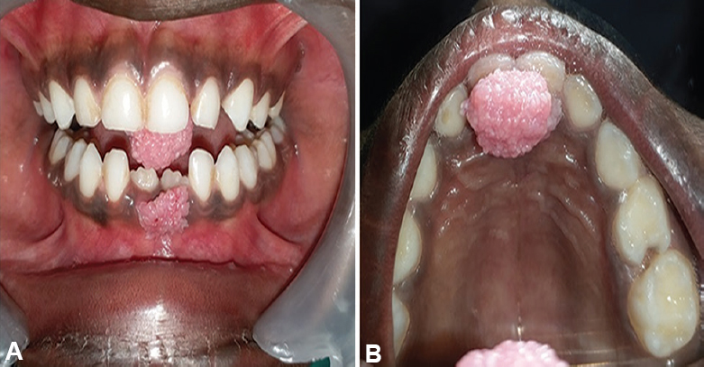
- Showing growth with finger-like projections present on anterior mandibular gingiva where the growth of maxilla is contacting (A). Occlusal view of the exophytic pedunculated growth over incisive papilla (B).
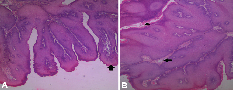
- Photomicrograph of haematoxylin and eosin (H and E)-stained section of maxillary growth in ×5 (A) showing multiple, exophytic finger-like projections (arrow) and ×10 (B) showing stratified squamous epithelium with hyperparakeratosis (arrowhead) and connective tissue core (arrow).
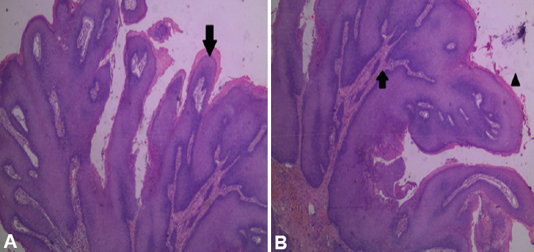
- Photomicrograph of haematoxylin and eosin (H and E)-stained section of mandibular growth in ×5 (A) showing multiple, exophytic finger-like projections (arrow) and ×10 (B) showing hyperplastic stratified squamous epithelium (arrowhead) with papillomatosis enclosing core of connective tissue (arrow).
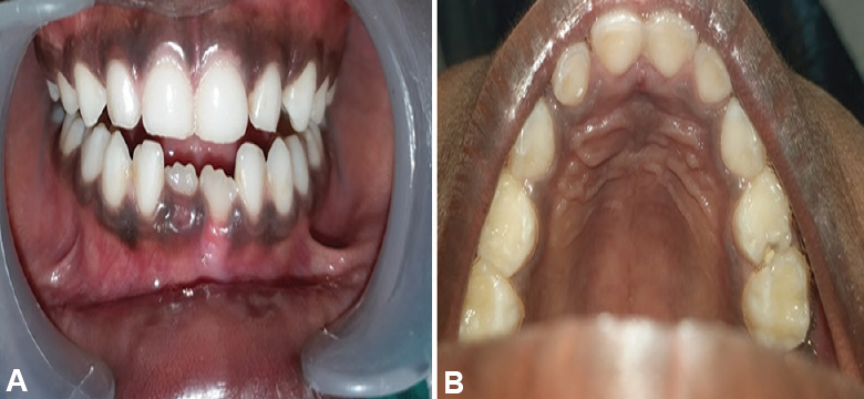
- A three month post-operative follow up image showing satisfactory healing and no recurrence in anterior mandibular gingiva (A) and over incisive papilla (B).
Acknowledgment
Authors acknowledge the department of Oral Pathology, Tamil Nadu Government Dental College and Hospital, Chennai, Tamil Nadu, India histopathological examination of tissue.
Conflicts of Interest: None.


