
Translate this page into:
Multiple cutaneous lesions in multiple myeloma responding to radiotherapy
*For correspondence: drmadhup1@gmail.com
-
Received: ,
This is an open access journal, and articles are distributed under the terms of the Creative Commons Attribution-NonCommercial-ShareAlike 4.0 License, which allows others to remix, tweak, and build upon the work non-commercially, as long as appropriate credit is given and the new creations are licensed under the identical terms.
This article was originally published by Wolters Kluwer - Medknow and was migrated to Scientific Scholar after the change of Publisher.
A 68 yr old male† patient presented to the department of Radiation Oncology, Dr. Ram Manohar Lohia Institute of Medical Sciences, Lucknow, India, in December 2012, with complaints of low back and right shoulder pain. Clinically multiple cutaneous lesions with bleeding points were seen (Fig. 1). X-ray of the shoulder revealed a large cutaneous lesion with no underlying bone destruction (Fig. 1C). Biopsy from the lesion (Fig. 1D, ×10 and Fig. 1E, ×20) revealed tumour positive for LCA (leucocyte common antigen) and CD138 while negative for cytokeratin (CK7), CK20, TTF1 (thyroid transcription factor 1), synaptophysin and cytokeratin AE1/AE3 suggestive of high-grade haematolymphoid malignancy of plasmablastic differentiation. Serum protein electrophoresis showed M band of IgG kappa. X-ray spine showed lytic lesions over the L3, L4, L5 and sacroiliac joints. Chemotherapy with bortezomib, cyclophosphamide and zoledronic acid was started. Radiotherapy (20 Gy in 5 fractions) with electrons (6 Mev) was delivered to right shoulder and the abdominal lesions. One month after delivery of radiotherapy, there was a partial response in cutaneous lesions present over the shoulder (Fig. 2A) and abdomen (Fig. 2B). At a follow up of 14 months, the patient continued to have partial response at the cutaneous sites and was continuing with chemotherapy and lost to further follow up.

- Cutaneous lesion on the (A) shoulder and (B) over abdomen. (C) X-ray showing cutaneous lesion with no bone destruction. Immunohistochemistry of the diopsy from the lesion (D) ×10 and (E) ×20 revealed tumour positive for LCA and CD138 while negative for CK7, CK20, TTF1, synaptophysin and AE1/AE3 malignancy with plasmablastic differentiation.
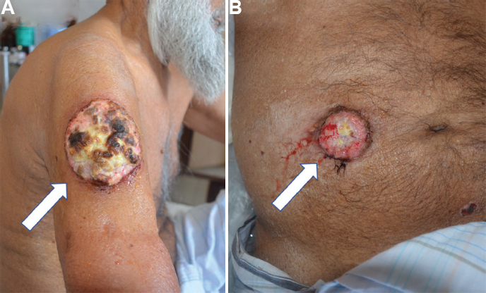
- Response to radiotherapy in (A) shoulder lesion and (B) abdominal cutaneous lesion.
Conflicts of Interest: None.


