
Translate this page into:
Melorheostosis: Dripping candle wax disease
*For correspondence: raju.vaishya@gmail.com
-
Received: ,
This is an open access journal, and articles are distributed under the terms of the Creative Commons Attribution-NonCommercial-ShareAlike 4.0 License, which allows others to remix, tweak, and build upon the work non-commercially, as long as appropriate credit is given and the new creations are licensed under the identical terms.
This article was originally published by Wolters Kluwer - Medknow and was migrated to Scientific Scholar after the change of Publisher.
A 29 yr old male†, presented in September 2019 to the departments of Orthopaedics & Joint Replacement Surgery, Indraprastha Apollo Hospitals, New Delhi, India, with pain and progressive deformity in the right foot (Fig. 1A) and leg (Fig. 1B), since 15 years. Radiographs revealed thickened cortices, with expanded bones and a pathognomic resemblance of 'dripping candle wax' appearance of the medial bones of the foot (Fig. 2A), ankle (Fig. 2B) and distal tibia (Fig. 2C), with arthritis of the midtarsal joints. The magnetic resonance imaging confirmed these findings (Fig. 3). A diagnosis of melorheostosis (Leri's disease) was made, which is a developmental disorder and can be diagnosed by radiographs due to its characteristic features. He was advised arthrodesis of the foot, but the patient was lost to follow up. Melorheostosis tends to follow a sclerotome distribution and can be monostotic or polystotic and often monomelic. It has a predilection for long bones, although it can be seen anywhere. Hands and feet may be frequently involved, however, the involvement of the axial skeleton is rare.
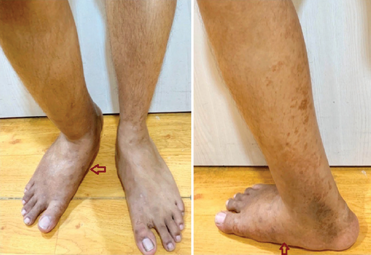
- Clinical photograph of the (A) foot, ankle, and (B) leg, showing deformity on the right side, with discolouration of the skin (arrows).
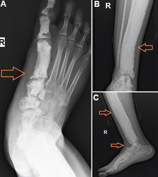
- Plain radiographs showing multi-osseous involvement of the (A) foot, (B) ankle, and (C) leg, bones, with cortical thickening, bone expansion and dripping candle wax appearance (arrows).
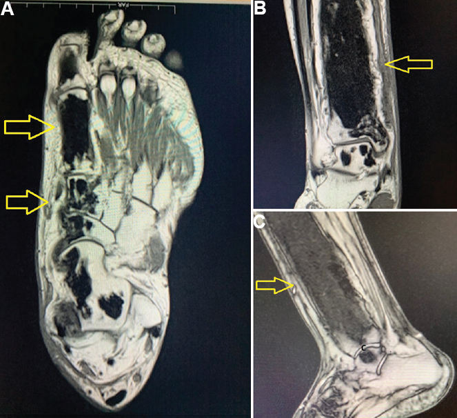
- Magnetic resonance images of the (A) foot and (B) ankle showing heterogeneous, sclerotic meta-diaphyseal expansion of the distal tibia. (C) Focal scleroitic areas are seen in the talus, calcaneus, navicular, cuboid, medial cuneiform, 1st metatarsal and phalanges of the big toe.
Conflicts of Interest: None.


