
Translate this page into:
Crippled woman with bilateral femur fracture
*For correspondence: paulguvera@gmail.com
-
Received: ,
This is an open access journal, and articles are distributed under the terms of the Creative Commons Attribution-NonCommercial-ShareAlike 4.0 License, which allows others to remix, tweak, and build upon the work non-commercially, as long as appropriate credit is given and the new creations are licensed under the identical terms.
This article was originally published by Wolters Kluwer - Medknow and was migrated to Scientific Scholar after the change of Publisher.
A 23 yr old female† presented to the department of Orthopedics, All India Institute of Medical Sciences (AIIMS), Bhubaneswar, India, on October 19, 2019 with a fracture of both femurs following a trivial fall 28 days back. The patient was completely bedridden and was put on skin traction till evaluated. Radiographs revealed pathological fracture of both femurs in the sub-trochanter region and punctate lesions in the skull (Fig. 1). Serum parathyroid hormone level was 1803 ng/dl (normal: 10-65 ng/dl); and serum alkaline phosphatase significantly elevated to 1031 U/l (normal: 100-290 U/l). Serum ionized calcium was 1.51 mmol/l (normal: 1-1.4 mmol/l) and serum phosphate was 2.0 mg/dl (normal: 2.5-5 mg/dl). Bone scan revealed increased uptake of the whole skeleton; polyostotic fibrous dysplasia. The diagnosis made was parathyroid adenoma with hyperparathyroidism leading to pathological fracture. Following confirmation of diagnosis, parathyroid glands were removed partially and serum calcium level stabilized. The patient was operated for fracture of the sub-trochanter region. The fracture in the right femur was fixed on December 23, 2018 and that of left femur on January 4, 2019 with an intramedullary nail as per anaesthetic fitness (Fig. 2A). Both femurs united well in the due course of time (Fig. 2B). Patient was prescribed calcium supplementation and high protein diet along with open chain and isotonic exercises. In a follow up after one year, the patient was leading a normal life with no recurrence.

- Fracture of the left and right femurs (yellow arrows), and punctate lesion of the skull (red arrows).
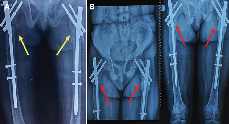
- (A) Immediate post-operative radiograph with implant in situ (yellow arrow) and (B) fracture completely healed at one year (red arrows).


