
Translate this page into:
Caseoma of the mitral annulus: A rare cardiac pseudotumour
* For correspondence: anantha25@yahoo.com
-
Received: ,
This article was originally published by Wolters Kluwer - Medknow and was migrated to Scientific Scholar after the change of Publisher.
A 72 yr old woman†, a known patient of diabetes mellitus with nephropathy for 15 years and end-stage renal disease on biweekly haemodialysis for a decade, was referred to Army Institute of Cardiothoracic Sciences, Pune, in January 2020, for echocardiographic evaluation. She complained of exertional dyspnoea (NYHA Class III) and easy fatigability with no angina, syncope, fever, weight loss or any embolic manifestations. Transthoracic echocardiography revealed concentric left ventricular hypertrophy with normal ejection fraction, dilated left atrium, grade II diastolic dysfunction and a well defined homogenous hyperechoic fixed mass in the posterior mitral leaflet, with severe mitral regurgitation without stenosis, with moderate pulmonary artery hypertension [PAH; right ventricular systolic pressure (RVSP)=40 mmHg]. Aortic valve was sclerosed with no stenosis or regurgitation (Figure A, B and Video 1). The differential diagnoses included mitral annular calcification (MAC), calcified thrombus, calcified vegetation, abscess and benign or malignant cardiac tumours. On cardiac magnetic resonance imaging, a focal lesion, hypointense on T1- and T2-weighted image was seen, measuring 22 × 20 mm, arising from the posterior mitral leaflet and adjoining annulus with no post-contrast enhancement (Figure C and D). These findings confirmed a caseoma, which is a chronic degenerative caseous calcification of the mitral annulus. The true incidence of this rare variant of MAC is unknown, with an estimated prevalence of 0.06 per cent in the general population and 2.7 per cent in the autopsy series of MAC. The current consensus is conservative medical management and avoiding surgical mitral valve replacement unless complicated by mitral valve dysfunction (mitral valve stenosis/regurgitation), systemic embolization (stroke) or uncertain diagnosis. She was unwilling for surgery and is presently being managed symptomatically at one year follow up with strict fluid compliance and adequate dialysis sessions. It is imperative for physicians to be aware of this benign condition and exclude other malignant mitral valve lesions. Surgery in such cases can be complicated by left ventricular perforation due to aggressive debridement and systemic embolization of the necrotic debris.
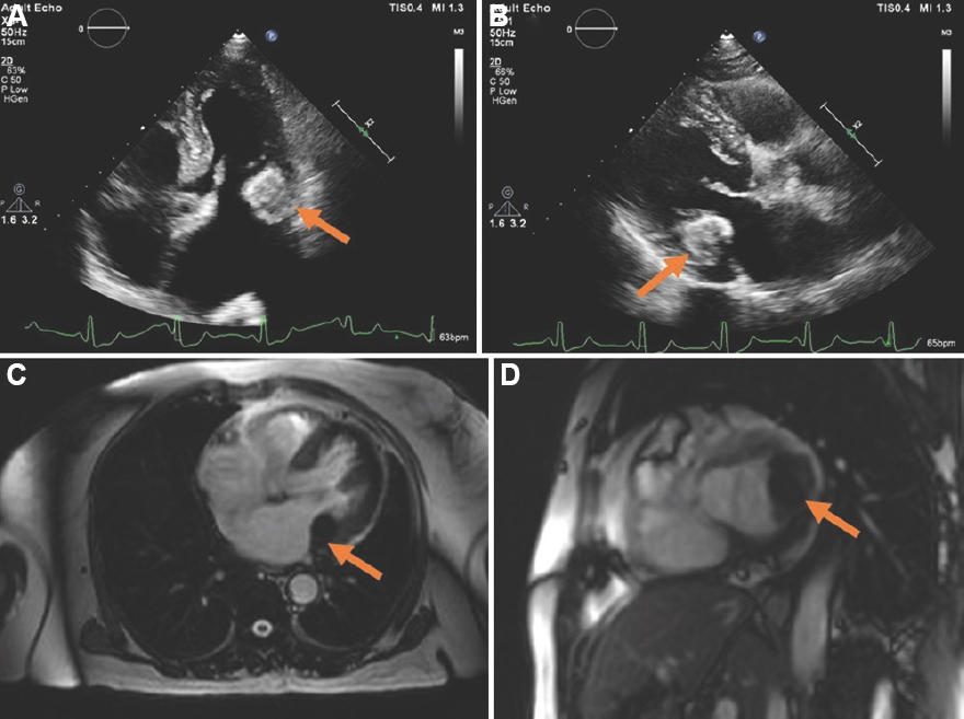
- (A) Apical four-chamber echocardiographic view showing well-defined homogenous hyperechoic mass from posterior mitral leaflet (arrow) with dilated left atrium. (B) Parasternal long-axis echocardiographic view showing calcified mitral leaflet (arrow). (C) T1-weighted magnetic resonance imaging in four-chamber view depicting hypointense focal lesion from mitral leaflet (arrow). (D) Short axis of magnetic resonance imaging showing calcified mitral leaflet and adjoining annulus (arrow).
Financial support and sponsorship
None.
Conflicts of interest
None.


