
Translate this page into:
A unique case of overlap syndrome with strange immunological findings
*For correspondence: dryashwant@ymail.com
-
Received: ,
This is an open access journal, and articles are distributed under the terms of the Creative Commons Attribution-NonCommercial-ShareAlike 4.0 License, which allows others to remix, tweak, and build upon the work non-commercially, as long as appropriate credit is given and the new creations are licensed under the identical terms.
This article was originally published by Wolters Kluwer - Medknow and was migrated to Scientific Scholar after the change of Publisher.
A 46 yr old female† presented to the outpatient department of Postgraduate Institute of Medical Education & Research, Chandigarh, India, in July 2019, with weakness and arthralgia for a duration of 2-3 months. Her peripheral blood sample was received in the department of Immunopathology for antinuclear antibody (ANA) testing. Indirect immunofluorescence ANA test (IIF-ANA) on human epithelial type 2 (HEp-2) cells showed a peculiar pattern in the form of haphazardly dispersed granules in both nucleus and cytoplasm (Figure A) and forming a zipper pattern within the mitosis (inset). IIF-ANA on composite section (rat liver, kidney tubules and gastric mucosa) showed classical anti-mitochondrial antibody (AMA) pattern in the form of granular positivity in the above tissues (Figure B-D, respectively). ANA and liver immunoblot thereafter were performed and found to be positive for CENP, AMA-M2 (Figure E and F) and SP100 autoantibodies (Figure F). The patient also had raised serum IgG, weak-positive p-ANCA and positive AMA-M2 on ELISA (110.0 U), whereas liver-kidney microsomes (LKM) and soluble liver antigen (SLA) were negative. On the basis of these immunoassay findings, the patient was given corticosteroid and ursodeoxycholic acid combination therapy. At three months follow up she remained asymptomatic.
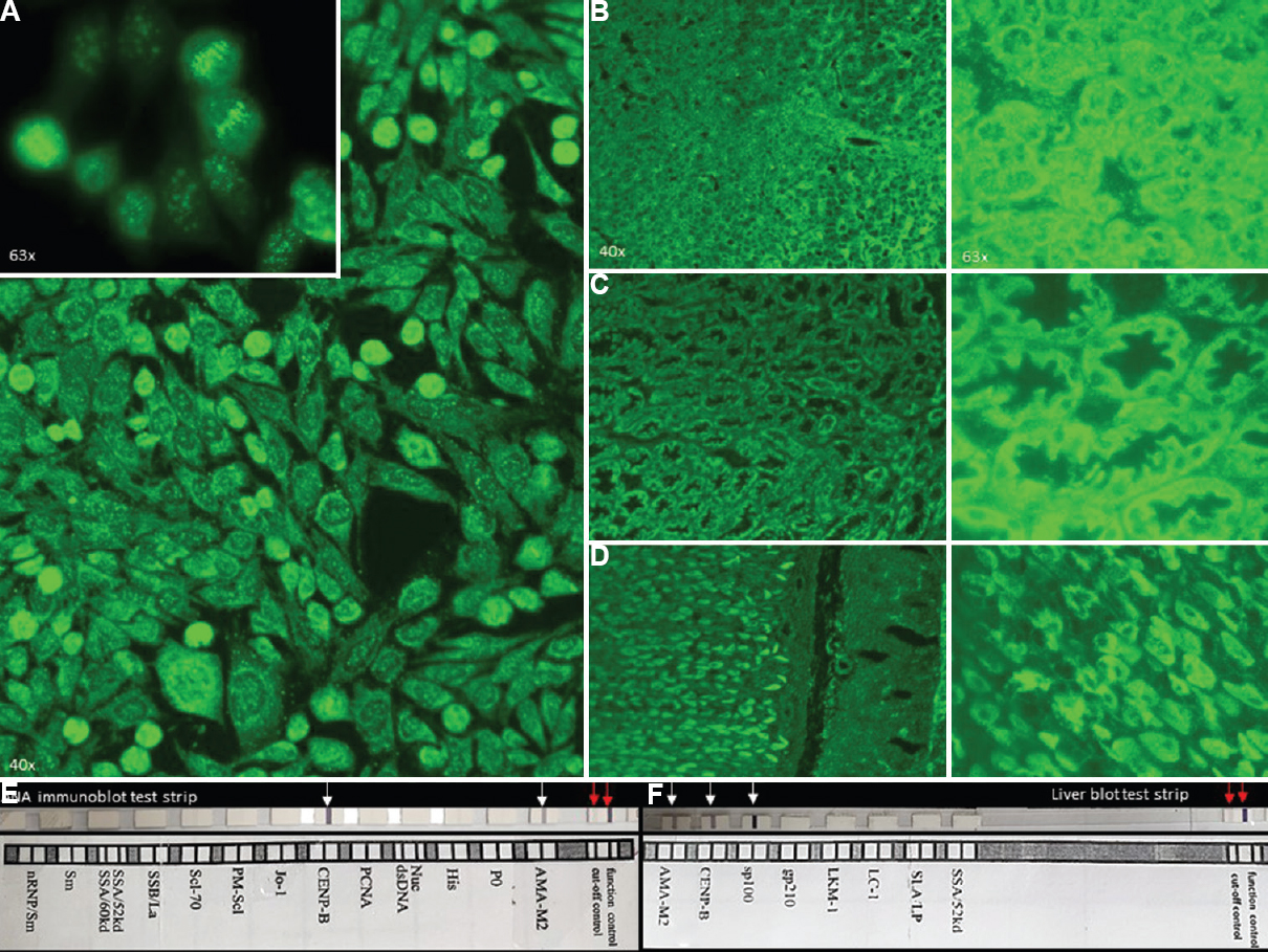
- (A) Indirect immunofluorescence antinuclear antibody (IIF-ANA) test on Hep-2 images showing a peculiar pattern in the form of haphazardly dispersed granules in both nucleus and cytoplasm and forming a zipper pattern within the mitosis (inset). (B-D) IIF-ANA test on composite section (rat liver, kidney tubules and gastric mucosa) showed classical anti-mitochondrial pattern in the form of granular positivity. (E) Antinuclear antibody and liver immunoblot are positive for CENPB (Centromere protein B), anti-mitochondrial-M2 (E and F, white arrow; red arrows are control bands) and (F) SP100 autoantibodies.
The above findings led to revised histopathological diagnosis of autoimmune hepatitis to overlap syndrome [autoimmune hepatitis - primary sclerosing cholangitis (AIH-PSC)]. This case is an example where a careful analysis of IIF-ANA on Hep-2 cells helped in correct diagnosis of the patient.
Conflicts of Interest: None.


