
Translate this page into:
A rare case of maxillary artery aneurysm
*For correspondence: drmlsaha@yahoo.com
-
Received: ,
This is an open access article distributed under the terms of the Creative Commons Attribution-NonCommercial-ShareAlike 3.0 License, which allows others to remix, tweak, and build upon the work non-commercially, as long as the author is credited and the new creations are licensed under the identical terms.
This article was originally published by Medknow Publications & Media Pvt Ltd and was migrated to Scientific Scholar after the change of Publisher.
A 44 yr old female presented to the department of General Surgery, Seth Sukhlal Karnani Memorial Hospital, Kolkata, India, in July 2015 with a slow growing painless mass in her left parotid region for the last 10 months which was not associated with fever or any constitutional symptoms. She denied any history of trauma to her face. There were no complaints of dysphagia, dysarthria or difficulty in opening mouth. She had no features of facial nerve palsy. There was no past history of surgery or medical illness. She was hypertensive and non-diabetic. On examination, a pulsatile globular swelling was found in her left parotid region (Fig. 1). Her neurological and cardiovascular examination was normal. An ultrasonography showed the presence of a hypervascular mass in the region of parotid, probably of arterial origin. A computed tomography angiography done subsequently revealed the presence of maxillary artery aneurysm along with synchronous intracranial aneurysms (Figs 2 & 3). She was referred to the cardiothoracic department for further management. However, she was lost to follow up. Maxillary artery aneurysms are exceedingly rare and most reported cases include pseudoaneurysms often occurring due to trauma or facial fractures. True aneurysms of maxillary artery are rare. Treatment includes coil embolization of the arteries.

- A globular swelling (9 cm × 8 cm) with smooth surface and regular margin in the left parotid region. The swelling has displaced her ear lobule backwards.
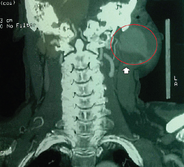
- Computed tomography angiogram of the neck showing a saccular aneurysm of the left maxillary artery involving the left masticator, parapharyngeal and infratemporal spaces. The red circle points out the maxillary artery aneurysm.
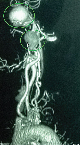
- Computed tomography angiogram (coronal cuts) of the same patient showing a concomitant left internal carotid artery aneurysm (green circles) along with the left maxillary artery aneurysm.


