
Translate this page into:
Cytology of disseminated cutaneous rhinosporidiosis
*For correspondence: nirmalyachakrabarti@gmail.com
-
Received: ,
This is an open access journal, and articles are distributed under the terms of the Creative Commons Attribution-NonCommercial-ShareAlike 4.0 License, which allows others to remix, tweak, and build upon the work non-commercially, as long as appropriate credit is given and the new creations are licensed under the identical terms.
This article was originally published by Wolters Kluwer - Medknow and was migrated to Scientific Scholar after the change of Publisher.
A 50 yr old male† farmer presented to the Surgery outpatient department, Burdwan Medical College, Burdwan, West Bengal, India, with soft, non-tender, swellings over multiple sites on the body (Fig. 1A and B) in February 2019, imparting a clinical diagnosis of soft tissue tumour. Fine-needle aspiration cytology showed plenty of endospores and sporangia of different sizes as intact (Fig. 2A and B) and ruptured (Fig. 3). Multiple punch biopsies showed thin epidermis and the presence of sporangia of different sizes in the dermis surrounded by histiocytes and lymphocytes and transepidermal elimination of the sporangium (Fig. 4A-C). The sporangia and endospores were periodic acid-schiff (PAS) positive confirming diagnosis of disseminated cutaneous rhinosporidiosis. The patient was operated for nasal rhinosporidiosis. After one week of dapsone therapy, the patient developed jaundice, haemolytic anaemia and skin rash. Dapsone was immediately discontinued following development of respiratory distress. Computed tomography thorax revealed multiple lung nodules with pleural effusion. Thoracentesis and treatment with cycloserine and ketoconazole failed to relieve symptoms even after two weeks following which the patient died.
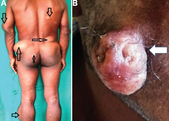
- (A) Patient with multiple swellings over the body. (B) Pedunculated swelling over the right cheek.
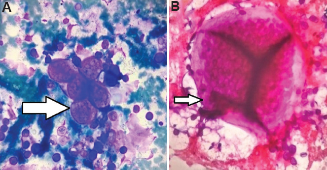
- (A) Cytology smear showing several endospores and growing sporangia of different sizes (Giemsa stain, ×40). (B) Mature sporangia (H and E stain, ×40).
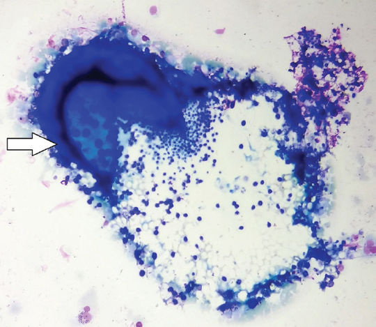
- Cytology smear showing ruptured mature sporangia liberating endospores (Giemsa stain, ×40).
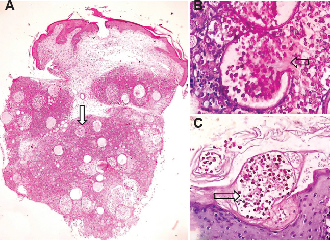
- (A) Punch biopsy of the facial lesion showing thinned out epidermis, growing sporangia in the deep dermis and within subcutaneous tissue, surrounded by dense lympho-histiocytic infiltrate (PAS stain, ×4). (B) A mature sporangia with a pore, through which endospores were seen to extrude in the mucoid matrix. (C) Transepidermal elimination of sporangia (PAS stain, ×40).
Conflicts of Interest: None.


