
Translate this page into:
Comparison of apoptotic bodies’ count & mitotic index in oral squamous cell carcinoma with regional lymph node involvement
For correspondence: Dr Noushin Jalayer Naderi, Department of Oral and Maxillofacial Pathology, Faculty of Dentistry, Shahed University, Tehran, Iran e-mail: jalayer@shahed.ac.ir
-
Received: ,
This article was originally published by Wolters Kluwer - Medknow and was migrated to Scientific Scholar after the change of Publisher.
Abstract
Background & objectives:
In early stages of oral cancers, 20-40 per cent of cases have occult metastasis in cervical lymph nodes. Biologic imbalance between cellular proliferation and death culminates in metastasis. The importance of cell cycle dysregulation in relation to lymph node involvement in oral squamous cell carcinoma (OSCC) has not been established yet. The aim was to determine the association between apoptotic bodies count and mitotic index in relation to regional lymph node involvement in OSCC.
Methods:
Thirty two methyl green-pyronin stained slides from paraffin-embedded sections of OSCC were evaluated for apoptotic bodies count and mitotic index in relation to regional lymph node involvement using light microscopy. Number of apoptotic bodies and mitotic figures were counted in 10 randomly selected hot spot areas (×400). Average count of apoptotic bodies and mitotic figures were determined and compared with regard to the presence/absence of lymph node involvement.
Results:
The count of apoptotic bodies in cases without metastasis to the regional lymph node was significantly higher than in cases with regional lymph node involvement. The mitotic index was not significantly different between groups in terms of regional lymph node involvement (P=0.24). No significant correlation was found between the apoptotic bodies count (r=−0.094, P=0.72) and mitotic index (r=−0.08, P=0.75) to the number of regional lymph nodes involved.
Interpretation & conclusions:
Based on the results, it is suggested that apoptotic cell count can be a good parameter for showing the possibility of regional lymph node involvement in people with OSCC who do not have clinical symptoms of lymph node involvement.
Keywords
Carcinoma
methyl green
oral
pyronin
squamous cell

In malignant transformation, apoptosis eliminates the cells with damaged DNA in response to certain stimuli such as radiation and anticancer drugs. When cells do not receive death (or apoptotic) signals, their number increases and cancer progresses. By examining apoptosis, the treatment process can be tracked1. Mitoses indicate the rapid proliferative activity of cells, however, these do not inevitably demonstrate the malignancy. But appearance of mitotic cells in epithelial tissue indicates a fast cellular turnover. The discrepancy between apoptosis and mitotic rate is marked by a pathological process which progresses towards malignancy2.
Squamous cell carcinoma is one of the most common malignancy of oral cavity3. The prognosis of oral squamous cell carcinoma (OSCC) is associated with the histopathologic grade of tumour, distant metastasis and involvement of the regional lymph nodes3. However, there is still no distinct protocol to predict the clinical course of OSCC from histopathologic specimens.
In the early stages of oral cancers, 20-40 per cent of cases have occult metastasis in cervical lymph nodes4. Both false positive and false negative results even in advanced diagnostic techniques such as positron emission tomography-computed tomography (PET-CT) lead to undertreatment or high extent dissection of cervical lymph nodes5. It is of practical importance to achieve a histopathologic method that can detect lymph node involvement without the need for high technique procedures.
Apoptotic counts may be a good way to predict the prognosis, but little is known about its association with metastasis. It has been shown that the apoptotic cells increased from premalignant lesions to OSCC6,7. Mitotic figures are more prevalent in dysplastic epithelium and malignant neoplasms compared to normal squamous epithelium due to higher rate of proliferation2.
The mitotic cells have been previously reported as a potentially good parameter to determine the fate of cancer cells8. An increasing rate of mitotic cells from normal epithelium to carcinoma and even to lower histopathologic grades of OSCC has been shown previously9-11. Since the dysregulation of the cell cycle contributes to carcinogenesis, it may also be associated with metastasis to lymph nodes.
Analyzing the proliferative activity and cellular death has both diagnostic as well as prognostic importance in detecting the risk of developing carcinoma12. However, efforts have not been made to solidify the importance of such a dual analysis of the apoptotic and mitotic cells in relation to lymph node involvement in OSCC. So, the aim of this study was to compare the apoptotic bodies’ count and mitotic index with respect to the regional lymph node involvement in OSCC.
Material & Methods
This study was retrospectively conducted at the department of Oral and Maxillofacial Pathology, Shahed University, Tehran, Iran, after it was approved by the Ethical Committee of Shahed University. The histopathologic and medical records of patients with OSCC were collected from the archive of Iran National Tumor Bank, Cancer Biology Research Center, Cancer Institute of Iran, Tehran University of Medical Sciences, Tehran, Iran, from 2005-2018. Considering d=0.05, Z=1.96 at 95 per cent confidence level of test and power of sample size=0.928 (Version 25; IBM Company, Chicago, IL, USA) based on the results of Seth and Agarwal13, the sample size was determined as 16 cases under each group.
Histopathology: The histopathologic slides of OSCC with adequate mass and good tissue fixation were included in the study. Subjects with incomplete medical and histopathologic records and history of radiotherapy/chemotherapy were excluded from the study. The marginal sections were not entered into the study. Haematoxilin-eosin-stained slides of all samples were examined using an optical microscope (ZEISS, Germany) at ×40 magnification. Samples with adequate tumoural bulk, without areas of massive necrosis and haemorrhage were selected. A total of 32 OSCC comprising two sets included 16 samples with and without regional lymph node involvement, respectively, were evaluated.
Formalin-fixed, paraffin-embedded sections (4 µm) of samples were first deparaffinized and then hydrated in distilled water, incubated in methyl green-pyronin solution (2% aqueous methyl green and 1% aqueous pyronin; Merck, Germany) for 2-7 min. Then, the sections were rinsed with distilled water, dipped in absolute alcohol and dried. Finally, after clearing in Xylene the sections were mounted. With methyl green-pyronin staining, RNA and DNA were stained by pyronin and methyl green and were seen in red and green colours, retrospectively14. Contracted cells with dense, sharply delineated chromatin within an eosinophilic cytoplasm were considered apoptotic bodies13. The apoptotic cells were spherical to oval which contain pyknotic nucleus (Fig. 1). The mitotic index was examined by counting the mitotic cells. The mitotic figures were identified based on van Diest et al15. The absence of the nuclear membrane, extensions of nuclear material and separate chromosome mass were considered as mitotic figures.
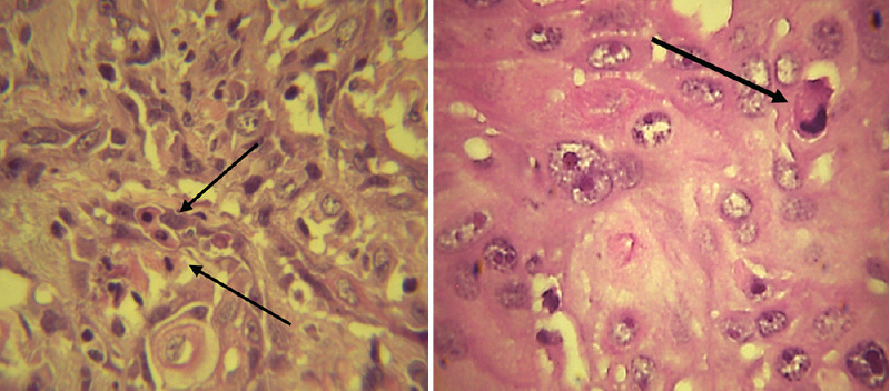
- Apoptotic cells (arrows) in oral squamous cell carcinoma (methyl green-pyronin, ×400).
The number of mitotic figures and apoptotic bodies were counted in 10 randomly selected hot spot areas at ×400 magnification. Average count of apoptotic bodies and mitotic figures were determined for each sample. The findings were evaluated and compared with regard to the presence or absence of lymph node involvement. Light microscopy (OLYMPUS BX40, Olympus Life Sciences, Tokyo, Japan) equipped with a digital camera (Sony ExWaveHAD, Model No. SSC-DC58AP; Tokyo, Japan) was used for counting.
Statistical methods: Mann-Whitney U test was employed to compare the means of groups at P<0.05. The correlation between apoptotic bodies count and mitotic index in relation to the regional lymph node involvement was examined using Spearman’s correlation coefficient at P<0.05. The SPSS statistical software package (Version 25; IBM Corp., Chicago, IL, USA) was used.
Results
Out of 32 cases, 15 (46.8%) were male and 17 (53.1%) were female. The mean age of cases was 56.7±17.21. The mean number of involved lymph nodes was 4.44±1.12. The mean of tumour size was 2.7±1.2 cm. Of the 32, 27 (84.4%) cases were from tongue, three (9.4%) from floor of the mouth and 2 (6.2%) from buccal and lip mucosa.
The mean number of apoptotic bodies in cases with and without lymph node involvement were 56.69±4.98 and 161.69±13.05, respectively. The mean mitotic index in cases with and without lymph node involvement were 11.13±2.50 and 8.56±2.35, respectively (Fig. 2). The Table shows the descriptive characteristics of the apoptotic bodies count and mitotic index in cases with and without lymph node involvement.
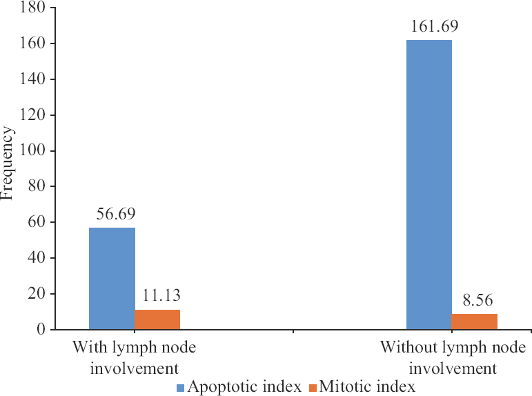
- Mean number of apoptotic bodies count and mitotic index in cases with and without lymph node involvement.
| Variables | Apoptotic bodies | Mitotic index | ||
|---|---|---|---|---|
| Mean±SEM | Median (minimum-maximum) | Meam±SEM | Median (minimum-maximum) | |
| With lymph node involvement | 56.69±4.98 | 58.5 (22-92) | 11.13±2.50 | 7 (0-33) |
| Without lymph node involvement | 161.69±13.05 | 171.5 (50-242) | 8.56±2.35 | 4.5 (0-31) |
SEM, standard error of mean
The Mann-Whitney U test revealed that apoptotic bodies count in cases without metastasis to regional lymph node was significantly higher than in cases with regional lymph node involvement. The mitotic index was not significantly different between groups in terms of regional lymph node involvement.
No significant correlation was found between the apoptotic bodies count (r=−0.094, P=0.72) and mitotic index (r=−0.08, P=0.75) to number of involved regional lymph nodes using the Spearman’s correlation coefficient test.
Discussion
The results show that the apoptotic bodies count in cases without metastasis to the regional lymph node was significantly higher than cases with regional lymph node involvement. The apoptotic bodies count and mitotic index were not correlated to the number of involved regional lymph nodes.
A lower rate of the apoptotic index has been related to increased tendency of lymph node involvement in OSCC12. This is consistent with the results of this study which showed a higher count of apoptotic bodies in cases without metastatic deposits of OSCC in regional lymph nodes as compared to cases with lymph node involvement. During an apoptotic process, the evacuated cells are killed. When malignant cells can escape the intrinsic and extrinsic pathways of cell death, then metastasis will occur. A poor apoptotic pathway reportedly contributes to carcinogenesis which has important outcomes in cancer treatment and prognosis16, a higher rate of apoptosis may be accompanied by a slower rate of tumour growth and lesser biological activity17. This is similar to the results of the present study which showed the association between apoptotic count and lymph node involvement.
The mitotic index was not significantly different between groups in terms of regional lymph node involvement. It has been shown that the rate of mitotic cells increased from normal epithelium to carcinoma and even to lower histopathologic grades of OSCC9-11. Although the previous findings suggest a possible association between mitosis and histopathologic grade, it cannot indicate the probability of lymph node metastasis. No literature was found about the association between the mitotic index and lymph node involvement in OSCC. Determining the relationship between quantities number of mitosis count and the likelihood of lymph node involvement requires further studies. The findings showed that the involvement of lymph nodes as well as the number of involved nodes did not correlate with mitotic cell counts. This finding suggests that the association between malignant tumour and metastasis is not just a simple relationship between malignant cell proliferation and death. It is important to note that the rate of apoptosis and proliferation regulate the cellular population2.
The distinction between apoptotic cells and pyknotic nucleus is difficult in conventional histochemical staining. Visual errors in detecting the apoptotic cells affects the histopathologic grading18. Haematoxylin-eosin does not represent a specific stain to display the DNA and RNA changes, so it cannot provide a valid, reliable interpretation of nucleus and nucleoli changes. Due to contradictory results on the relationship between apoptotic bodies’ count and prognosis, haematoxylin-eosin has not been patented until now19-21.
Methyl green is specifically linked to DNA and pyronin to RNA, accordingly, it reveals the nuclei of replicated cells, undifferentiated non-proliferative nuclei and degenerated DNA22. It has been shown that methyl green-pyronin had a good outcome in assessing the nucleus and nucleoli changes in premalignant lesions23. This approach provides a simple and cost-effective way to detect the cellular objects such as apoptotic bodies in light microscopy. The present study showed that the methyl green-pyronin had a satisfactory result in displaying the cellular changes. Recognizing the apoptotic cells was easier and reduces the possibility of confusing the apoptotic bodies with small dyskeratotic cells. This is cosimilar to the finding of Sumedha et al6 who showed that the methyl green-pyronin-staining was a simple method to detect apoptotic cells in routine histopathologic examination. In the present study too, using methyl green-pyronin stained slides provided more accurate, reliable results as compared to the routine stain of hematoxylin-eosin under light microscopy. The main limitation of this study was the small sample size, since some samples had to be excluded because of insufficient tumoural tissue and lack of clinical information.
Based on these findings, it is suggested that the apoptotic bodies count can be used as a reliable parameter in determining the lymph node involvement in cases without clinical evidence of cervical lymph node involvement even in small incisional biopsies of OSCC. To determine the prognosis of SCC, different factors such as tumour stage, lymphovascular and perineural invasion have been used. Since examination of cellular changes on histopathologic slides is the first step in the diagnosis of a malignant lesion, the present study assessed the role of apoptotic bodies and mitotic count in association with regional lymph node involvement in OSCC. Further studies are recommended to determine the cut-off point of the apoptotic bodies count in association with regional lymph node involvement.
Financial support and sponsorship
The study was financially supported by the research grant from the Shahed University. Biological materials were provided by the Iran National Tumor Bank which was funded by Cancer Institute of Tehran University of Medical Sciences, for Cancer Research.
Conflicts of interest
None.
Acknowledgment:
The authors acknowledge the team of Iran National Tumor Bank, Cancer Biology Research Center, Cancer Institute of Iran, Tehran University of Medical Sciences, for their assistance in archive retrieval and preparing the histopathologic slides.
References
- Apoptosis in cancer: From pathogenesis to treatment. J Exp Clin Cancer Res. 2011;30:87.
- [Google Scholar]
- Robbins &cotran pathologic basis of disease (10th ed). Philadelphia: Elsevier; 2021.
- Grade as a prognostic factor in oral squamous cell carcinoma: A population-based analysis of the data. Laryngoscope. 2014;124:688-94.
- [Google Scholar]
- Predictive factors of occult neck metastasis in patients with oral squamous cell carcinoma. Braz J Otorhinolaryngol. 2016;82:543-7.
- [Google Scholar]
- Imaging evaluation of lymphadenopathy and patterns of lymph node spread in head and neck cancer. Expert Rev Anticancer Ther. 2015;15:207-24.
- [Google Scholar]
- A histochemical comparison of methyl green-pyronin, and hematoxylin and eosin for detecting apoptotic cells in oral squamous cell carcinoma, oral leukoplakia, oral submucous fibrosis and normal oral mucosa. Biotech Histochem. 2015;90:264-9.
- [Google Scholar]
- Apoptosis in normal epithelium, premalignant and malignant lesions of the oropharynx and oral cavity: A preliminary study. Eur J Cancer B Oral Oncol. 1995;31B:380-3.
- [Google Scholar]
- Apoptosis, mitosis, PCNA and bcl-2 in normal, leukoplakic and malignant epithelia of the human oral cavity: Prospective, in vivo study. Oral Oncol. 1997;33:419-25.
- [Google Scholar]
- Evaluation of mitotic figures and cellular and nuclear morphometry of various histopathological grades of oral squamous cell carcinoma: Comparative study using crystal violet and Feulgen stains. Sultan Qaboos Univ Med J. 2018;18:e149-54.
- [Google Scholar]
- Correlation of mitosis obtained by using 1% crystal violet stain with Ki67LI in histological grades of oral squamous cell carcinoma. J Oral Biol Craniofac Res. 2018;8:234-40.
- [Google Scholar]
- Evaluation of efficacy of 1% Crystal Violet &Nuclear Fast Red stain compared to Haematoxyline &Eosin stain for assessing mitotic figures in oral premalignant and malignant lesions. J Exp Ther Oncol. 2016;11:165-70.
- [Google Scholar]
- Apoptosis in premalignant and malignant squamous cell lesions of the oral cavity: A light microscopic study. Indian J Pathol Microbiol. 2009;52:164-6.
- [Google Scholar]
- Apoptotic count as a guide for histological grading of carcinoma esophagus: A light microscopic study. J Lab Physicians. 2009;1:11-4.
- [Google Scholar]
- Expression of cell cycle and apoptosis-related proteins in ameloblastoma and keratocystic odontogenic tumor. Ann Diagn Pathol. 2013;17:518-21.
- [Google Scholar]
- Reproducibility of mitosis counting in 2,469 breast cancer specimens: Results from the Multicenter Morphometric Mammary Carcinoma Project. Hum Pathol. 1992;23:603-7.
- [Google Scholar]
- Apoptosis and prognosis in cancer: Rationale and relevance. J R Coll Surg Edinb. 2000;45:211-9.
- [Google Scholar]
- Potential molecular prognostic markers in head and neck squamous cell carcinomas. Head Neck. 2001;23:147-59.
- [Google Scholar]
- Lymphnode metastasis in head and neck squamous cells carcinoma: Multivariate analysis of prognostic variables. J Exp Clin Cancer Res. 1999;18:79-83.
- [Google Scholar]
- Prognostic significance of apoptosis in squamous cell carcinoma of oral cavity with special reference to TNM stage, histological grade and survival. Indian J Pathol Microbiol. 2001;44:257-9.
- [Google Scholar]
- Comparison of staining of mitotic figures by haematoxylin and eosin-and crystal violet stains, in oral epithelial dysplasia and squamous cell carcinoma. Indian J Dent Res. 2007;18:101-5.
- [Google Scholar]
- Methyl green-pyronin Y staining of nucleic acids: Studies on the effects of staining time, dye composition and diffusion rates. Biotech Histochem. 2003;78:27-33.
- [Google Scholar]
- Detection of nucleoproteins in squamous cell carcinoma, and dysplastic and normal mucosa in the oral cavity by methyl green-pyronin staining. J Oral Sci. 2010;52:239-43.
- [Google Scholar]


