
Translate this page into:
Unilateral submandibular gland agenesis
-
Received: ,
This is an open access article distributed under the terms of the Creative Commons Attribution-NonCommercial-ShareAlike 3.0 License, which allows others to remix, tweak, and build upon the work non-commercially, as long as the author is credited and the new creations are licensed under the identical terms.
This article was originally published by Medknow Publications & Media Pvt Ltd and was migrated to Scientific Scholar after the change of Publisher.
A 46 yr old female came to the ENT department of Sree Balaji Medical College and Hospital, Chennai, India, with minimal painless swelling in the bilateral cheek and left submandibular region for two months, in September 2014. The patient had no significant medical history other than multiple carious tooth extractions. Bimanual palpation could not locate the submandibular gland on the right side but revealed a prominent submandibular gland on the left side. Contrast-enhanced computed tomography (CT) scan of neck revealed bilateral prominent parotid glands with no focal lesions. Right submandibular gland was absent. Left submandibular gland, seen in normal location, was mildly enlarged with no focal lesions (Figs 1 & 2). Diagnosis of right submandibular gland agenesis with compensatory hypertrophy of contralateral submandibular gland and both parotid glands was made. The patient was called for follow up after four months, but there was no clinically significant change in the swelling. Submandibular gland agenesis is an uncommon disorder and may be hereditary/syndromic/sporadic and unilateral/bilateral. Unilateral submandibular gland aplasia is usually asymptomatic and discovered incidentally during imaging. Clinical features may include mouth dryness, chewing difficulty and dental caries. Clinicians as well as radiologists must be aware of this condition to reassure the patient. Treatment is primarily supportive and aims at reducing xerostomia and its effects, if present.
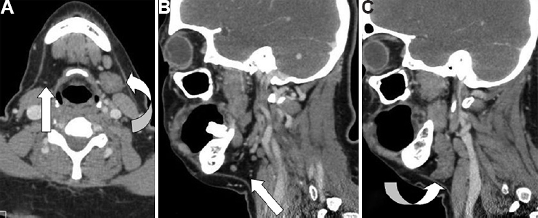
- (A) Axial contrast-enhanced computed tomography (CECT) of neck shows absent right submandibular gland (white arrow) in right submandibular fossa and hypertrophied left submandibular gland (curved arrow). (B) Sagittal reformat of CECT shows absent right submandibular gland in right submandibular fossa (white arrow). (C) Sagittal reformat of CECT shows hypertrophied left submandibular gland (curved arrow).
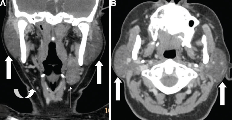
- (A) Coronal reformat of contrast-enhanced computed tomography (CECT) of neck shows hypertrophied bilateral parotid glands (white arrow), absent right submandibular gland in right submandibular fossa (curved arrow) and hypertrophied left submandibular gland (thin white arrow). (B) Axial CECT shows hypertrophied bilateral parotid glands (white arrow).
Acknowledgment
Author acknowledges the help received from Dr Hari MS, Department of ENT, Sree Balaji Medical College and Hospital, Chennai.


