Translate this page into:
Plexiform neurofibromatosis type 1
* For correspondence: sksharma.aiims@gmail.com
This is an open access article distributed under the terms of the Creative Commons Attribution NonCommercial ShareAlike 3.0 License, which allows others to remix, tweak, and build upon the work non commercially, as long as the author is credited and the new creations are licensed under the identical terms.
This article was originally published by Medknow Publications & Media Pvt Ltd and was migrated to Scientific Scholar after the change of Publisher.
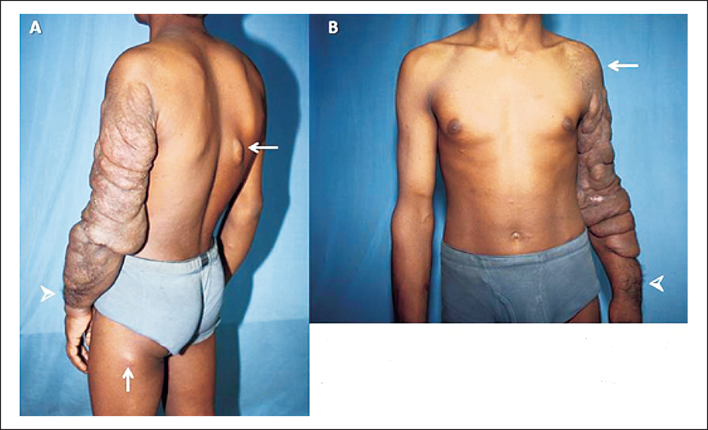
A 16 yr old boy presented to the department of Medicine, All India Institute of Medical Sciences (AIIMS), New Delhi, India, in 2013 with primary cosmetic concern about swelling in the left upper limb which was progressively increasing over the last few years. A rigorous clinical work-up revealed presence of several café-au-lait macules (CALM) (>5) along with multiple neurofibromas (Fig. A arrows), plexiform neurofibroma of the left upper limb, hypertrichosis (Fig. A & B arrowheads) and hyperpigmentation (Fig. B arrow). Magnetic resonance imaging (MRI) of the adrenals, brain and spine was non-contributory. There was no cognitive impairment, signs of skeletal deformities, ocular and acoustic abnormalities or any family history of neurofibromatosis. A diagnosis of plexiform neurofibromatosis type 1 was made which is an autosomal dominant disorder. Plexiform neurofibroma has been classically described as a “bag of worms”. Amongst the complications, malignant transformation although rare, has been reported and due to progressive increase in size of the plexiform neurofibroma, disfigurement and bony erosions can occur. No definitive treatment was offered at this time. However, a regular follow up was advised keeping in mind the risk of future complications such as hypertension, etc.