
Translate this page into:
Vein finder – A cost-effective tool to screen pneumothorax in neonatal intensive care unit
*For correspondence: mail4shajin@gmail.com
-
Received: ,
This is an open access journal, and articles are distributed under the terms of the Creative Commons Attribution-NonCommercial-ShareAlike 4.0 License, which allows others to remix, tweak, and build upon the work non-commercially, as long as appropriate credit is given and the new creations are licensed under the identical terms.
This article was originally published by Wolters Kluwer - Medknow and was migrated to Scientific Scholar after the change of Publisher.
A two day old neonate† presented to the Neonatal Intensive Care Unit, department of Paediatrics, Makunda Christian Leprosy and General Hospital, Assam, India, during the month of September 2019. He was depressed at birth, was on ventilatory support and had sudden spontaneous desaturation. A vein finder (Fig. 1) was used, which showed an obvious pneumothorax (Fig. 2), which was confirmed with chest X-ray. Due to this timely intervention, the neonate was saved.
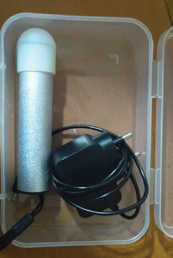
- A vein finder device.

- A vein finder device being used to screen a baby with pneumothorax.
The vein finder makes use of infrared light which helps to screen for pneumothorax. Placing the detector on the neonate's chest produces transillumination, which aids in detection.
Pneumothorax is a common condition in the neonatal intensive care unit (NICU). Fibre-optic cold light source is commonly used to screen for pneumothorax in the NICU. Although pneumothorax requires an X-ray, when there is a tension pneumothorax, there may not be enough time to wait for an X-ray, as it is an emergency. Furthermore, a cold light source may be expensive for smaller NICUs, but having a screening device such as a vein finder, which is cheap, helps to intervene quickly. Using this device in our sick newborn care unit, especially in resource-limited settings, can save a lot of neonates.
Conflicts of Interest: None.


