
Translate this page into:
Tuberculous optochiasmatic arachnoiditis & myeloradiculopathy
*For correspondence: imranrizvi09@gmail.com
-
Received: ,
This is an open access journal, and articles are distributed under the terms of the Creative Commons Attribution-NonCommercial-ShareAlike 4.0 License, which allows others to remix, tweak, and build upon the work non-commercially, as long as appropriate credit is given and the new creations are licensed under the identical terms.
This article was originally published by Wolters Kluwer - Medknow and was migrated to Scientific Scholar after the change of Publisher.
A 12 yr old boy† presented to the Neurology outpatient department, King George's Medical University, Lucknow, India, in August 2019, with complaints of fever and headache, paraparesis and bilateral vision loss of 12 month duration. On examination, the patient was fully conscious, vision was 6/60 in both the eyes and optic atrophy was noted bilaterally. Motor examination revealed lower motor neuron paraparesis. Cerebrospinal fluid examination revealed lymphocytic pleocytosis, raised protein and low sugar. The Xpert Mycobacterium tuberculosis/rifampicin assay (Cepheid, USA) was positive. Magnetic resonance imaging (MRI) brain showed multiple tuberculomas in the optochiasmatic region (Fig. 1). MRI spine showed syrinx in the cervical spine, cerebral spinal fluid loculations around lumbar spine, clumping and enhancement of roots suggestive of arachnoiditis (Fig. 2). The patient was treated with anti-tuberculosis drugs along with corticosteroids; after one month of follow up, he showed improvement in fever and headache although there was no improvement in his vision or lower limb strength. This case represents classical complications of tuberculous meningitis which can leave a patient chronically disabled.
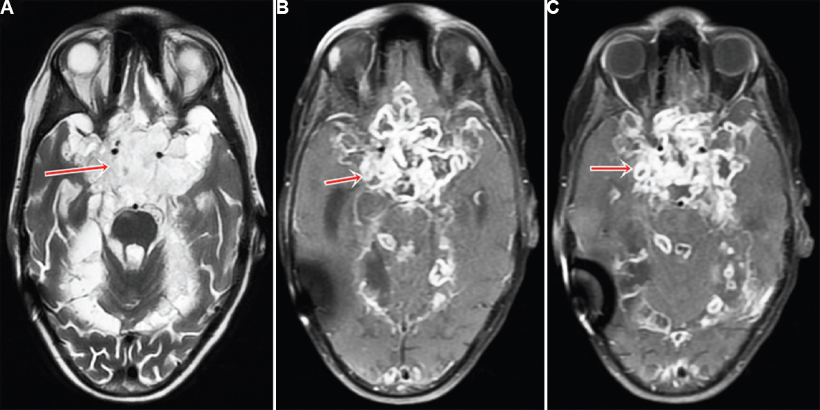
- (A) T2-weighted magnetic resonance image of brain showing multiple lesions of varying intensity in the optochiasmatic region (red arrow). (B & C) Post-contrast T1 image showing multiple peripherally enhancing lesions in the optochiasmatic area (red arrow).
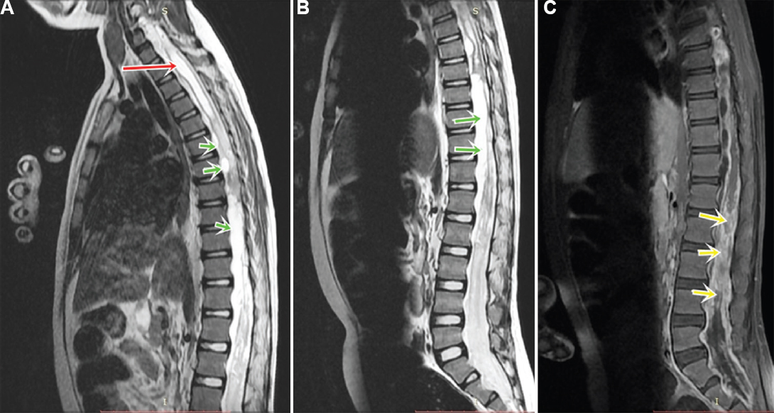
- (A) Magnetic resonance imaging (MRI) cervicodorsal spine showing syrinx involving the cervical spine (red arrow) and cerebral spinal fluid loculations along the dorsal spine (green arrows). (B) MRI lumbosacral spine T2-weighted image showing cerebral spinal fluid loculations around lumbar spine (green arrows). (C) Post-contrast image showing enhancement and clumping of roots (yellow arrows).
Conflicts of Interest: None.


