
Translate this page into:
Tuberculosis of the shoulder: ‘Caries sicca’
* For correspondence: dr_vipulvijay@yahoo.com
-
Received: ,
This is an open access journal, and articles are distributed under the terms of the Creative Commons Attribution-NonCommercial-ShareAlike 4.0 License, which allows others to remix, tweak, and build upon the work non-commercially, as long as appropriate credit is given and the new creations are licensed under the identical terms.
This article was originally published by Medknow Publications & Media Pvt Ltd and was migrated to Scientific Scholar after the change of Publisher.
A 22 yr old male presented to the Orthopaedics Outpatient Department of Indraprastha Apollo Hospital, New Delhi, India, in June 2015 with gradually progressive painful swelling and restricted movements in the left shoulder since last five months. Local examination revealed muscle wasting as well (Fig. 1A, arrow). The radiograph (Fig. 1B) showed the typical Phemister's triad (lysis in greater tuberosity of humerus, perilesional osteopenia and reduced joint space). Magnetic resonance imaging revealed soft-tissue collection in the subacromial and subdeltoid bursae and marrow oedema in humeral head (Fig. 2A, arrow). The erythrocyte sedimentation rate (ESR) was 65 mm/h, and C-reactive protein was 14.26 ng/ml. Aspiration revealed thick, yellow coloured, caseous material, histopathologically consistent with tuberculosis with the presence of Langhans giant cells and epithelioid granuloma (Fig. 2B and C, inset).
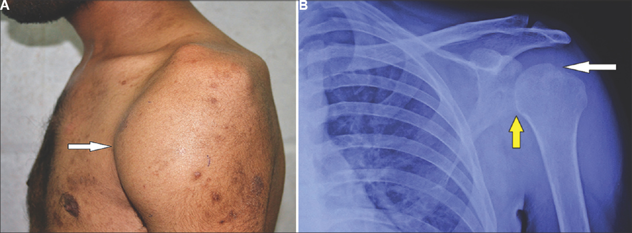
- (A) Clinical photograph of the patient depicting soft-tissue swelling on the anterior aspect of the shoulder with significant wasting of muscles around the shoulder (arrow). (B) Anteroposterior radiograph of the shoulder showing the Phemister's triad - lytic lesion of the greater tuberosity (white arrow), diffuse periarticular osteopenia and joint space reduction (yellow arrow).
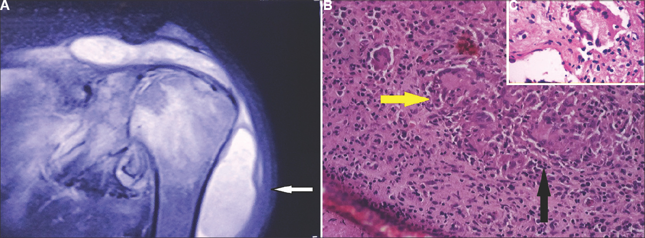
- (A) T2 magnetic resonance image of the shoulder with a large subacromial and subdeltoid collection with marrow oedema in the humeral head (arrow). (B) Photomicrograph of the aspirate depicting the typical Langhans-type giant cells (yellow arrow) with epithelioid granuloma (black arrow) (H & E, ×10). (C) (Inset) An enlarged view of the photomicrograph showing the isolated granuloma with the giant cell having the nucleus in typical horseshoe shape (H & E, ×30).
Pain and swelling responded to anti-tubercular therapy (three months of isoniazid, rifampicin, pyrazinamide and ethambutol followed by six months of isoniazid and rifampicin) with an improved range of motion. The local radiograph also showed improved osteopenia and decreased soft-tissue shadow (Fig. 3).
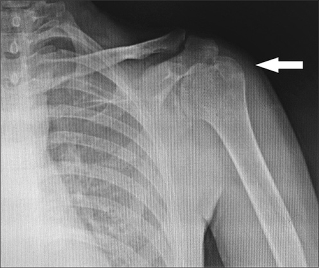
- Post-treatment anteroposterior radiograph of the shoulder with improved osteopenia and reduction of the soft-tissue shadow around the shoulder as compared to the original radiograph (arrow).


