
Translate this page into:
‘SpaceX-sign’ in presumed solitary circumscribed retinal astrocytic proliferation
* For correspondence: drmohitdogra@gmail.com
-
Received: ,
This article was originally published by Wolters Kluwer - Medknow and was migrated to Scientific Scholar after the change of Publisher.
A 45 yr old asymptomatic male† presented to the department of Ophthalmology, Postgraduate Institute of Medical Education & Research, Chandigarh, in August 2018, for a routine eye examination. The best corrected visual acuity in the right eye was 6/6 and the left eye was 6/9. The right eye had a normal fundus while the left eye showed a yellowish-white, pearl-shaped lesion, nasal to the foveal center (Figure A). The swept source optical coherence tomography (SS-OCT) scan through the lesion revealed a well demarcated, hyper-reflective lesion arising from the outer retina and retinal pigment epithelium (RPE) without involvement of the inner retina. The tapered appearance of the lesion (in SS-OCT imaging) resembled the superior part of the ‘SpaceX’ space shuttle (Figure B). The clinical examination and SS-OCT were suggestive of presumed solitary circumscribed retinal astrocytic proliferation (p-SCRAP). The patient was counselled about the benign course of the lesion and advised regular follow up. p-SCRAP generally occurs in middle-aged males and is solitary, circumscribed and well defined retinal lesions. The exact origin of p-SCRAP lesion (due to fibrous metaplasia of the RPE cells or retinal glial cells) is debatable. Vision and the size of the lesion remain stable in most of the cases. Less than 25 cases of this entity have been previously reported in the literature. p-SCRAP needs to be differentiated from other lesions such as retinal astrocytoma, astrocytic hamartoma, retinoblastoma, myelinated nerve fibres, granulomas, choroidal neovascular membrane and reactive gliosis. To conclude, p-SCRAP is a rare and unusual clinical entity, and SS-OCT imaging is indispensable to make the correct diagnosis.
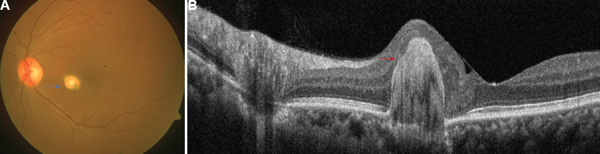
- (A) Fundus photograph of the left eye showing a yellowish-white, pearl-shaped lesion (blue arrow), nasal to the foveal centre. (B) Swept source optical coherence tomography scan showing a hyper-reflective lesion involving the outer retina and retinal pigment epithelium with sparing of inner retinal layers, the tapered appearance of the lesion (red arrow) resembles the superior part of ‘Space-X’ space shuttle.
Financial support and sponsorship
None.
Conflicts of interest
None.


