
Translate this page into:
Sirolimus for multifocal infantile haemangiomas with hepatic & adrenal involvement
*For correspondence: jhumaji@gmail.com
-
Received: ,
This is an open access journal, and articles are distributed under the terms of the Creative Commons Attribution-NonCommercial-ShareAlike 4.0 License, which allows others to remix, tweak, and build upon the work non-commercially, as long as appropriate credit is given and the new creations are licensed under the identical terms.
This article was originally published by Wolters Kluwer - Medknow and was migrated to Scientific Scholar after the change of Publisher.
Multifocal haemangiomas are a rare subtype of infantile haemangiomas (IHs) with multiple complications. A 40 day old male infant† with multifocal IH presented to the Emergency department at the All India Institute of Medical Sciences, New Delhi, India, in January 2019, with eye involvement, liver failure and high-output cardiac failure (Fig. 1A). The child was initially managed with propranolol to which the lesions were refractory. Angiogenesis inhibitor sirolimus was tried with good clinical response. The lesions regressed in size (Fig. 1B) and liver functions improved after two weeks of therapy. Unfortunately, the child succumbed to a nosocomial infection at four weeks of intensive care unit stay. Post-mortem examination revealed multifocal haemangiomas in the liver (Fig. 2A) and adrenal gland (Fig. 2B). The histopathology confirmed IHs (Fig. 3). This case highlights the challenges faced by the treating team in managing multifocal haemangiomas with hepatic involvement. Sirolimus appears to be a promising therapy in these cases.
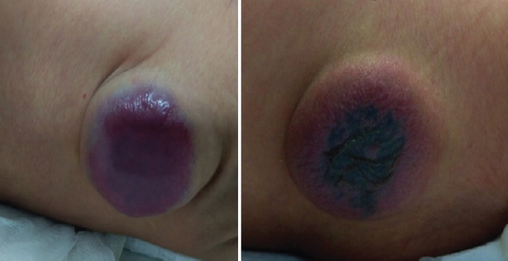
- (A) Lesion over the lower back before sirolimus therapy. (B) Image taken at three weeks after sirolimus therapy (notice reduced size and fading of the lesion).
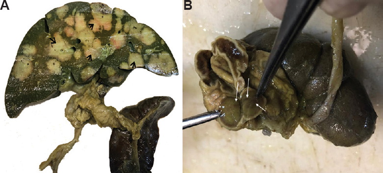
- (A) The gross specimen of the liver showing multiple varying sized sub-capsular grey-white lesions with central dark brown areas (arrows) with areas of congestions all over the liver. (B) The gross specimen of the adrenal gland showing similar varying sized grey-white lesions (arrows).

- Histopathology of liver showing intra-hepatic vascular tumour (arrows), comprising cleft-like areas, lined with endothelial cells. A few entrapped benign bile ducts were seen within the lesion (A, ×100). The endothelial cells (arrows) lining the vascular spaces did not show atypia (B, ×200) and were positive for CD34 stain (arrows) (C, ×200). Similar vascular lesion (black arrows) was also identified inside the left adrenal gland (blue arrows) (D, ×40), and the lesional cells (black arrows) were positive for CD34 stain (E, ×40). In skin also, similar lesions were identified (F, ×100).
Acknowledgment:
Authors acknowledge the contributions of Drs Viswanath, Sagar Tugal and Rakesh Lodha from the department of Pediatrics and Dr Hena Khandakar from the department of Pathology in the case management and for providing images.
Conflicts of Interest: None.


