
Translate this page into:
Sign from Sister Mary Joseph’s nodule
*For correspondence: cjj13390530038@126.com
-
Received: ,
This article was originally published by Wolters Kluwer - Medknow and was migrated to Scientific Scholar after the change of Publisher.
A 54 yr old† man was admitted to general surgery department of Jinhua Municipal Central Hospital, PR China, with a two-week history of cutaneous plaque developed on his umbilicus in March 2018. A tan, swelling and indurated nodule around the umbilicus was found (Fig. 1). He was diagnosed with ascending colon carcinoma and accepted laparoscopic radical operation six weeks ago. Pathology of the resected specimens showed a poorly differentiated adenocarcinoma. Histopathological finding of the cutaneous biopsy showed infiltration of the dermis by differentiated adenocarcinoma cells (Fig. 2). A diagnosis of Sister Mary Joseph’s nodule was made, the lesion in the umbilicus was caused by metastases from the colon carcinoma. After detailed condition assessment (K-ras: wild type), intravenous (IV) chemotherapy was administered every two weeks [oxaliplatin 85 mg/m2 ivgtt, day 1; leucovorin 400 mg/m2 ivgtt, day 1; 5-FU 400 mg/m2 IV bolus on day1, then 1200 mg/m2/day × two days (total 2400 mg/m2 over 46-48 h) continuous infusion; cetuximab 500 mg/m2 ivgtt, day 1, for 12 cycles]. No tumour progression was found at one year follow up.
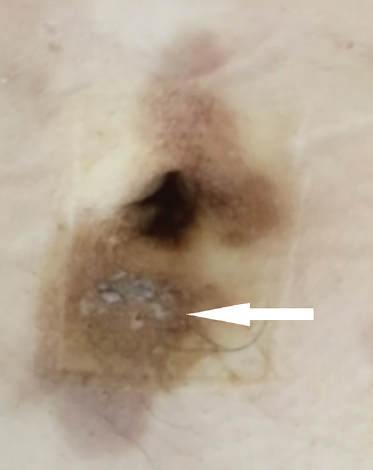
- An ill-defined, firm, indurated plaque in and around the umbilicus (arrow) was found on examination.
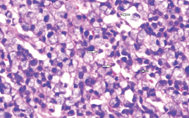
- Hematoxylin & eosin staining of skin biopsy sample showing extensive infiltration of the dermis by poorly differentiated adenocarcinoma cells (arrows, ×400).
Conflicts of Interest: None.


