
Translate this page into:
Segmental arterial mediolysis; vascular it is, not vasculitis
*For correspondence: dr.guptaranjan@gmail.com
-
Received: ,
This is an open access journal, and articles are distributed under the terms of the Creative Commons Attribution-NonCommercial-ShareAlike 4.0 License, which allows others to remix, tweak, and build upon the work non-commercially, as long as appropriate credit is given and the new creations are licensed under the identical terms.
This article was originally published by Wolters Kluwer - Medknow and was migrated to Scientific Scholar after the change of Publisher.
A 48 yr old man† presented to the department of Rheumatology, All India Institute of Medical Sciences, New Delhi, India, in September 2017 with painful swellings over the left elbow and chest for six months with intermittent dyspnea (Figure A). Chest radiograph showed miliary mottling of the lungs prompting initiation of anti-tubercular therapy, but he continued to develop new swellings with increase in size of existent ones. These were fusiform arterial aneurysms on computed tomography angiography (Figure B). Coronary arteriography done for progressively worsening dyspnea showed beaded appearance of the left coronary artery (Figure C), which was against the diagnosis of mycobacterial mycotic aneurysms. Systemic vasculitis was unlikely in view of normal inflammatory markers and negative serological tests. Therefore, non-inflammatory vasculopathy was strongly considered. Excision and bypass grafting for impending rupture of brachial aneurysm revealed segmental arterial mediolysis (SAM) on histopathology (Figure D and E). However, the patient succumbed due to catastrophic hemorrhage from another aneurysm. SAM is an important non-inflammatory cause for vascular aneurysms that can rarely involve the coronary arteries.
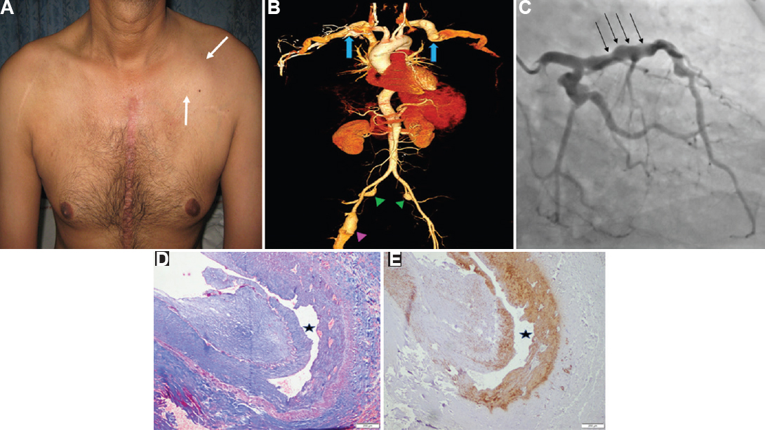
- (A) Clinical photograph showing chest wall swelling (white arrow). (B) Computed tomography arteriography volume rendered image demonstrating aneurysmal bilateral subclavian (blue arrows), internal iliac (green arrowheads) and right femoral arteries (pink arrowhead). (C) Coronary angiogram demonstrates beaded appearance of left coronary artery (black arrows). (D) Histopathological examination of the excised brachial aneurysm (H and E, ×200) and (E) smooth muscle actin (IHC, ×200), respectively, showing pathognomonic dissection in the arterial media (black star).
Conflicts of Interest: None.


