
Translate this page into:
‘Sea-fan’ neovascularization: Unusual presentation of proliferative diabetic retinopathy
*For correspondence: ramanuj.samanta@gmail.com
-
Received: ,
This article was originally published by Wolters Kluwer - Medknow and was migrated to Scientific Scholar after the change of Publisher.
A 50 yr old type-2 diabetic maleϯ, sub-optimally controlled on oral hypoglycaemic agents for 10 years, presented to the department of Ophthalmology, All India Institute of Medical Sciences, Rishikesh, India, in November 2019. He complained of blurred vision for three months. Best-corrected visual acuity was 6/18 in the right eye (RE) and 6/36 in the left eye (LE). Anterior segment was unremarkable. Dilated fundus examination revealed moderate non-proliferative diabetic retinopathy (DR) in the RE and proliferative DR with ‘Sea-fan’ neovascularization (SFN) along superior arcade and macular oedema in the LE (Figs 1A and B). He underwent pan-retinal photocoagulation in the LE and bilateral intravitreal anti-vascular endothelial growth factor (anti-VEGF) injections. Follow up after nine months showed nuclear sclerosis cataract and partially regressed SFN with lasered proliferative DR in the LE (Figs 2A and B). SFN is classically reported in sickle-cell retinopathy and rarely with thrombocytosis, sarcoidosis and retinitis pigmentosa. Laboratory investigations for sickle-cell disease and sarcoidosis were negative in this case. This report highlights SFN as an atypical presentation of proliferative DR, which might have occurred due to chronic ischaemia following microvascular occlusion and consequent raised VEGF level.
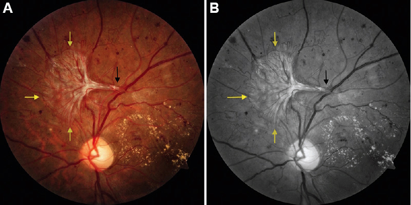
- Fundus image (A) and red-free photograph (B) of left eye showing a large frond of Sea-fan neovascularization (yellow arrows) with a fleshy stalk originating from major retinal vessel (black arrow). Multiple small neovascularizations and circinate ring of hard exudates are also seen.
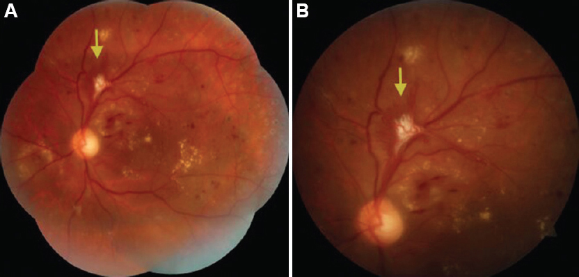
- Montage fundus photograph (A) and enlarged superior fundus field (B) of the left eye at follow up showing peripheral laser scars and partial regression of Sea-fan neovascularization (yellow arrow).
Conflicts of Interest: None.


