
Translate this page into:
Scapular mass in a young man with renal failure
*For correspondence: kohlihs2009@gmail.com
-
Received: ,
This is an open access journal, and articles are distributed under the terms of the Creative Commons Attribution-NonCommercial-ShareAlike 4.0 License, which allows others to remix, tweak, and build upon the work non-commercially, as long as appropriate credit is given and the new creations are licensed under the identical terms.
This article was originally published by Wolters Kluwer - Medknow and was migrated to Scientific Scholar after the change of Publisher.
A 26 yr old male†, on irregular haemodialysis for nine months presented to the department of Nephrology, Postgraduate Institute of Medical Education & Research, Chandigarh, India, in February 2019, with a three-month history of swellings around both the shoulder joints. These were gradually increasing in size but were painless without causing any movement restriction. Examination was notable for a multilobular mass over each of the shoulder joints and the scapulae, of non-tender, and firm consistency with no increased vascularity. Similar masses were observed over the bilateral heads of femur. Laboratory tests revealed serum calcium of 10 mg/dl (normal: 8.4-10.2 mg/dl), serum phosphate of 6.8 mg/dl (normal: 2.5-4.5 mg/dl), 25-hydroxy-vitamin D3 of 35 ng/ml (normal: 30-50 ng/ml) and intact parathormone of 880 pg/ml (normal: 15-65 pg/ml, up to nine times the upper limit is considered normal in end-stage renal failure). Radiography revealed dense chunks of calcification over the scapulae, glenohumeral joints and acetabulofemoral joints bilaterally (Figure A-C). Ultrasonography of the neck revealed no parathyroid adenoma. Needle aspiration from the scapular masses showed calcific deposits on Von Kossa's stain. Diagnosis of secondary tumoral calcinosis due to secondary hyperparathyroidism was made. The patient was started on sevelamer bicarbonate (800 mg thrice a day), and dialysis (low calcium dialysate) thrice a week. At the last follow up (6 months), laboratory parameters normalized and the swellings subsided.
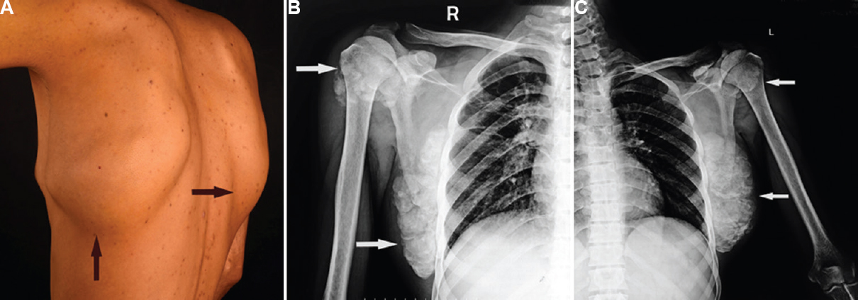
- (A) Bilateral scapular masses (arrows). (B) Chunks of calcification over the right glenohumeral joint and scapula (arrows). (C) Chunks of calcification over the left glenohumeral joint and scapula (arrows).
Conflicts of Interest: None.


