Translate this page into:
Quantitative assessment of the elasticity values of liver with shear wave ultrasonographic elastography
Reprint requests: Dr Nazan Ciledag, Ankara Oncology Research & Education Hospital, Department of Radiology 06460, Demetevler, Ankara/Turkey e-mail: drnazangokbayrak@yahoo.com.tr
-
Received: ,
This is an open-access article distributed under the terms of the Creative Commons Attribution-Noncommercial-Share Alike 3.0 Unported, which permits unrestricted use, distribution, and reproduction in any medium, provided the original work is properly cited.
This article was originally published by Medknow Publications & Media Pvt Ltd and was migrated to Scientific Scholar after the change of Publisher.
Abstract
Background & objectives:
Tissue stiffness in liver is related to tissue composition, which is changed by cirrhosis, hepatocellular carcinoma or metastases. Shear wave ultrasonographic elastography is a new imaging technique by which the elasticity of soft tissue can be measured quantitatively. The aim of this study was to measure the elasticity values of liver segments in healthy volunteers.
Methods:
One hundred twenty seven healthy volunteers (89 women, 38 men; mean age 37, 72 ± 9.11 yr, range 17-63 yr) were examined on shear wave elastography and ultrasonography by using convex probe with a frequency of 3 MHz. Individuals with liver hepatosteatosis, cirrhosis, chronic liver disease, or focal liver lesions were excluded from the study.
Results:
The mean elasticity values of right posterior, right anterior, left medial and left lateral segments of the liver was determined as 4 (±2.2), 3.3 (±2.1), 3.8 (±2.1), and 3.7 (±1.9) kPa for each segments, respectively. There was no significant difference in liver elasticity values between men and women.
Interpretation & conclusions:
In this preliminary study the elasticity values of liver segments were measured by shear wave ultrasonographic elastography in normal healthy volunteers. Further studies, comparing elasticity values of normal and pathologic tissues are needed to detect the diagnostic role of this new technique.
Keywords
Elasticity
elastography
liver
shear wave ultrasoun

Measurement of in vivo elasticity of liver can be a valuable, non invasive test to quantify tissue stiffness of liver, for the diagnosis of nonalcoholic steatohepatitis and prediction of liver fibrosis. Shear wave elastography is a method based on acoustic radiation force as a compression source to induce subtle tissue displacement1. The tissue displacements generated by acoustic push pulse used in shear wave elastography is related to tissue stiffness, while tissue recovery response is related to tissue viscoelastic properties2. In addition, shear wave elastography is positively correlated with the major mechanical properties of tissue indicating material rigidity3. Although acoustic radiation force induced transient elastography has been used for delineating tissue structure via mechanical properties in numerous applications including chronic liver diseases45, only a few studies have reported on shear wave elastographic ultrasound imaging of liver6.
A new diagnostic imaging technique called as conventional or dynamic elastography based on uniform, external compression reveals the physical properties of soft tissue by characterizing the differences in stiffness between the region of interest and the surrounding. Dynamic elastography is subjective qualitative, operator dependent method with low reproducibility, and does not give true elasticity values in kPA78. Dynamic ultrasonographic elastography has already been shown to be useful for the differential diagnosis of cancers in the breast91011, thyroid12 and prostate1314 as well as for lymph node15 characterization. Shear wave ultrasonographic elastography is a recently developed, non invasive method for measuring tissue elasticity, which gives a local assessment at each point of interest of an organ in kilopascals (kPa)17. This imaging method is operator-independent, reproducible, and quantitative. It has been used with success in the evaluation of breast lesions10.
To facilitate the widespread clinical use of the shear wave elastography, the elasticity values of normal soft tissues of liver must be established, so that the effect of steatohepatitis or liver fibrosis on the elasticity of the affected tissue can be investigated relative to baseline healthy liver tissue. So far, only one study has presented in vivo values for liver18. Therefore, the aim of this study was to assess the quantitative elasticity values of different liver segments in healthy volunteers.
Material & Methods
Study population: Between May to August 2010, 127 healthy volunteers from radiology or radiation oncology staff (89 women, 38 men; mean age: 37.72 ± 9.11 yr, range: 17-63 yr) were examined using both shear wave elastography and supersonic ultrasonography in Ankara Oncology Research and Education hospital, Ankara, Turkey. Cases with liver hepatosteatosis, cirrhosis, chronic liver disease, or focal liver lesions were excluded from the study. Also, individuals with body mass index higher than 30 kg/m2 were excluded from the study due to the limitation of shear wave ultrasonographic imaging of lesions deeper than 5 cm. The study group included volunteers who were followed with ultrasonography and serum biochemical tests routinely on yearly basis for at least for five years and both ultrasonographic examination and biochemical tests were normal in all volunteers.
The following parameters were recorded for each volunteer at the time of the study: age, sex, weight, height, and body mass index. The Ankara Oncology Research and Education hospital ethics review board approved this study, and informed written consent was obtained from all volunteers.
Ultrasound and shear wave ultrasonographic elastography technique: Shear wave ultrasonographic elastography of the different segments of liver was performed after a dedicated ultrasonography examination by using a 3 MHz covex transducer (Supersonic Imaging System, France). The right lobes of the patients were examined in left lateral decubitus, with the right arm in maximum abduction. The left lobes were examined in supine position, with right and left arm in maximum abduction. Scanning was performed with minimal scanning pressure applied by the operator, while the patients were asked to stop breathing for a moment, in order to minimize breathing motion. All measurements were performed by the same radiologist and reported in kilopascals. The tip of the transducer was covered also 5 mm of ultrasound gel and placed on the skin smoothly without compressing the tissue. Shear wave ultrasonographic elastography is based on the automatic generation and analysis of transient shear waves. This method uses the acoustic radiation force of the ultrasound wave to push the tissue instead of using a compression force (called stress), as is used in conventional dynamic ultrasonographic elastography. The acoustic displacement of tissue is free of user dependence and is reproducibile (Fig. 1).
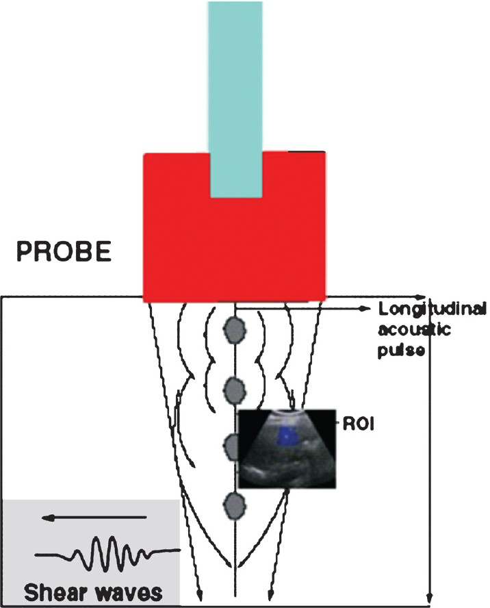
- Schematic image showing physical principles of ultrasonographic shear wave elastography. Transmission of longitudinal acoustic pulse leads to tissue displacement, which results in propagation of shear waves away from region of interest (ROI) with ultrasound tracking beams lateral to single push beam.
Statistical analysis: Statistical analyses were performed by using SPSS software system (version 15.0, Chicago, IL). Pearson correlation analysis and t-test for independent samples were used for data analysis. Descriptive statistics were used to summarize the characteristics of the subjects including the means and SDs of all continuous variables.
Results & Discussion
The mean elasticity values for the right posterior, right anterior, left medial and left lateral segments of the liver were determined as 4 kPa ±2.2 kPa (range:1-15), 3.9 kPa±2.1 (range:1-13) kPa, 3.8 kPa±2.1 (range:1-13) kPa, and 3.7 kPa±1.9 (range 1-12) kPa for each segments, respectively. There was no significant difference in liver elasticity values between men and women. No correlation was observed between age, and the elasticity values of the liver segments.
Assessment of liver fibrosis and determining the level of fibrosis are important in hepatology, to guide the antifibrotic action of different treatments. In clinical practice, the following three methods are used for the diagnosis and evaluation of the liver fibrosis: (i) liver biopsy, which is stil considered to be “gold Standard”; (ii) serological markers of fibrosis and their mathematical combination which is suggested in recent years to be an alternative method to biopsy; and (iii) transient elastography which is a new, simple and non invasive method used to measure liver stiffness in kPa19. Transient elastography is based on the progression of an elastic shear wave within the liver. It allows liver stiffness measurements which enables the assessment of liver disease severity, using a 1-dimensional ultrasound transducer and receiver mounted on the same axis as a vibrator, producing a low-frequency pulse or shear wave. When the probe tip is placed perpendicularly against the skin between the ribs overlying the liver and triggered, the rate of progression of the shear wave is measured. The major advantage of shear wave elastographic ultrasonography over transient elastography is the capability of real-time imaging of various target size and target positions. Transient elastography utilizes a vibration at limited range and target tissues without imaging support as a 10mm wide and 40 mm long cylinder, which is required to lie between 25 and 65 mm below the skin and free from large vascular structures. The area available for transient elastography is limited to a portion of right lobe, and practically it is difficult to compare the stiffness at different parts of the liver. In this study shear wave ultrasonographic measurements were performed in four different segments at the same time point. Foucher et al20 reported that transient elastography frequently failed to compute liver stiffness in patients with massive ascites or body mass index over 28 kg/m2, and real-time ultrasonographic shear wave measurements showed a similar tendency.
Shear wave ultrasonographic elastography is a novel imaging technique that has been proposed as an alternative, rapid, easy, non invasive method to assess liver elasticity. It uses the acoustic radiation force and acoustic pulses can be focused at different depths in the tissue at supersonic speed and are enhanced by forming a Mach cone, which increases the shear wave propagation. The Young's modulus reflects the speed of shear wave propagation and is directly related to the tissue elasticity values, reported in kilopascals, shown on a real time colour-coded elastography map of the region of interest (Fig. 2). The major advantages of the shear wave ultrasonographic elastography are the operator independency, reproducibility, higher spatial resolution, and the ability to perform a quantitative evaluation of elasticity values without manual compression artifacts.

- Left liver lobe elasticity map obtained by using shear wave ultrasonographic elastography in 43 yr old male volunteer. The elasticity score was 2.69kPa.
Osaki et al6 reported the correlation between shear wave velocity values and both serum biochemical parameters (such as albumin, hyaluronic acid) and liver fibrosis stage6. They have reported that, the average shear wave velocity for nonalcoholic steatohepatitis was 1.34 ± 0.26. Horster et al21 evaluated the correlation between the transient elastography stiffness values and liver fibrosis stages. They have reported that valsalva manoeuvre did not significantly alter shear wave elasticity value and variance.
In conclusion, in this preliminary study the elasticity values of liver segments were measured by shear wave ultrasonographic elastography in healthy volunteers. The major limitation of this study was non availability of elasticity values in pathologic tissues. Further studies need to be conducted in large number of patients comparing elasticity values between normal and pathologic tissues to detect the diagnostic role of this new technique.
References
- Shear wave elasticity imaging: a new ultrasonic technology of medical diagnostics. Ultrasound Med Biol. 1998;24:1419-35.
- [Google Scholar]
- Acoustic radiation force impulse imaging: a parametric analysis of factors affecting image quality. Proc IEEE Ultrasonics Symposium 2003
- [Google Scholar]
- A finite-element method model of soft tissue response to impulsive acoustic radiation force. IEEE Trans Ultrason Ferroelectr Freq Control. 2005;52:1699-712.
- [Google Scholar]
- Performance of a new elastographic method (ARFI technology) compared to unidimensional transient elastography in the noninvasive assessment of chronic hepatitis C. Preliminary results. J Gastrointestin Liver Dis. 2009;18:303-10.
- [Google Scholar]
- Evaluation of acoustic radiation force impulse elastography for fibrosis staging of chronic liver disease: a pilot study. Liver Int. 2010;30:538-45.
- [Google Scholar]
- Shear wave velocity is a useful marker for managing nonalcoholic steatohepatitis. World J Gastroenterol. 2010;16:2918-25.
- [Google Scholar]
- Sonoelastic determination of human skeletal muscle elasticity. J Biomech. 1995;28:1145-54.
- [Google Scholar]
- Realtime elastography. A new ultrasound procedure for the reconstruction of tissue elasticity. Radiologe. 2003;43:850-5.
- [Google Scholar]
- Breast disease: clinical application of US elastography for diagnosis. Radiology. 2006;239:341-50.
- [Google Scholar]
- Breast lesions: quantitative elastography with supersonic shear imaging - preliminary results. Radiology. 2010;256:297-303.
- [Google Scholar]
- Elastography of breast lesions: initial clinical results. Radiology. 1997;202:79-86.
- [Google Scholar]
- Freehand ultrasound elastography of breast lesions: clinical results. Ultrasound Med Biol. 2001;27:1461-9.
- [Google Scholar]
- Initial experiences with real-time elastography guided biopsies of the prostate. J Urol. 2005;174:115-7.
- [Google Scholar]
- Differential diagnosis of malignant cervical lymph nodes at real-time ultrasonographic elastography and Doppler ultrasonography. Hung Radiol Online. 2010;6:10-3.
- [Google Scholar]
- Shear wave elastography: A new ultrasound imaging mode for the differential diagnosis of benign and malignant thyroid nodules. J Clin Endocrinol Metab. 2010;95:5281-8.
- [Google Scholar]
- Shear wave speed recovery in transient elastography and supersonic imaging using propagating fronts. Inverse Problems. 2006;22:681-706.
- [Google Scholar]
- Prevalence and factors associated with failure of liver stiffness measurement using FibroScan in a prospective study of 2114 examinations. Eur J Gastroenterol Hepatol. 2006;18:411-2.
- [Google Scholar]
- Comparing acoustic radiation force impulse imaging to transient elastography to assess liver stiffness in healthy volunteers with and without valsalva manoeuvre. Clin Hemorheol Microcirc. 2010;46:159-68.
- [Google Scholar]


