
Translate this page into:
Pulmonary arterio-venous malformation
*For correspondence: uttamkpatnaik@gmail.com
-
Received: ,
This is an open access journal, and articles are distributed under the terms of the Creative Commons Attribution-NonCommercial-ShareAlike 4.0 License, which allows others to remix, tweak, and build upon the work non-commercially, as long as appropriate credit is given and the new creations are licensed under the identical terms.
This article was originally published by Wolters Kluwer - Medknow and was migrated to Scientific Scholar after the change of Publisher.
A 16 yr old male child† presented to the department of Cardiology, Srirama Chandra Bhanja Medical College & Hospital, Cuttack, India, in July 2019, with complaints of dyspnoea and bluish discolouration of skin since eight years. Physical examination revealed central cyanosis and grade-3 clubbing (Fig. 1). The chest radiograph showed a rounded opacity in the left lung with a slight increase in size over three years (Fig. 2A and B). Routine echocardiography was normal (Fig. 3A). Contrast echocardiography with agitated saline showed delayed right-to-left shunt. After the injection of agitated saline, there was transfer of contrast bubbles from right heart chambers to the left heart after three cardiac cycles (Fig. 3B and C). There was a gradual build-up of contrast in the left heart while diminishing from the right heart (Fig. 3D and Video). Based on the classical clinical presentation, X-ray findings and echocardiographic evidence of delayed right-to-left shunt, a diagnosis of pulmonary arteriovenous malformation was made. This was confirmed by computed tomography scan. The patient was pending surgical intervention.
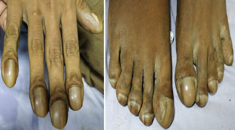
- Grade-3 clubbing with parrot-beak appearance of the fingers and toes.
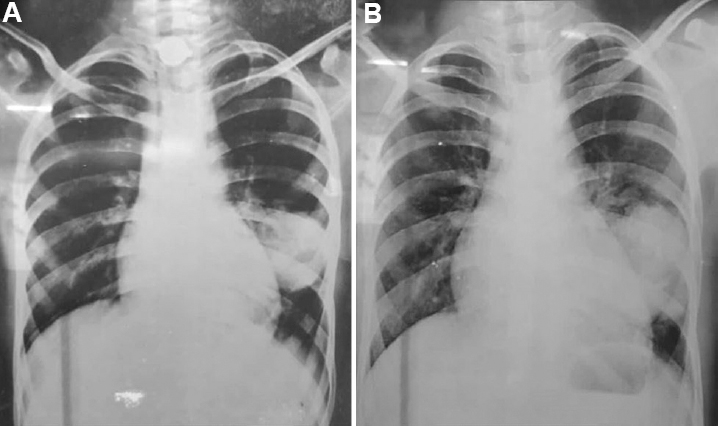
- Chest radiograph in postero-anterior projection showing homogenous rounded opacity in left lower zone. (A) Image taken in 2016 and (B) Image taken at presentation in 2019. The opacity persisted over a gap of three years with slight increase in size.
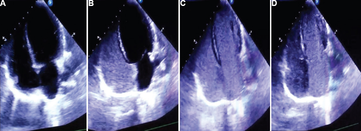
- Transthoracic echocardiographic images: (A) the normal cardiac chambers, septa and valves. (B) Image of saline contrast echocardiograph after injection of agitated saline, the contrast bubbles are seen in the right chamber of the heart. (C) Image after three cardiac cycles, where the bubbles appear in the left chamber of heart and (D) Further build-up of contrast bubbles in the left chamber while diminishing from the right chamber.
This case highlights the importance of diagnosing a rare case based on the typical presentation and using cost-effective, non-invasive imaging modalities in resource-limited settings lacking access to computed tomography scan.
 Video available at ijmr.org.in.
Conflicts of Interest: None.


