
Translate this page into:
Palatal fibroma in a geriatric patient: A rarity
*For correspondence: drsjayachandranmds@yahoo.com
-
Received: ,
This is an open access journal, and articles are distributed under the terms of the Creative Commons Attribution-NonCommercial-ShareAlike 4.0 License, which allows others to remix, tweak, and build upon the work non-commercially, as long as appropriate credit is given and the new creations are licensed under the identical terms.
This article was originally published by Wolters Kluwer - Medknow and was migrated to Scientific Scholar after the change of Publisher.
A 63 yr old male† patient reported to the department of Oral Medicine and Radiology, Tamil Nadu Government Dental College & Hospital, Chennai, India, in June 2019, with complaints of replacement of missing teeth. The patient gave a history of growth in the mid-palate for the past 20 years. Intraoral examination revealed a growth measuring 1.5 × 1 cm, brownish-black in colour, oval in shape with smooth surface. On palpation, growth was firm in consistency and non-tender with sessile base in the mid-palate (Fig. 1A).
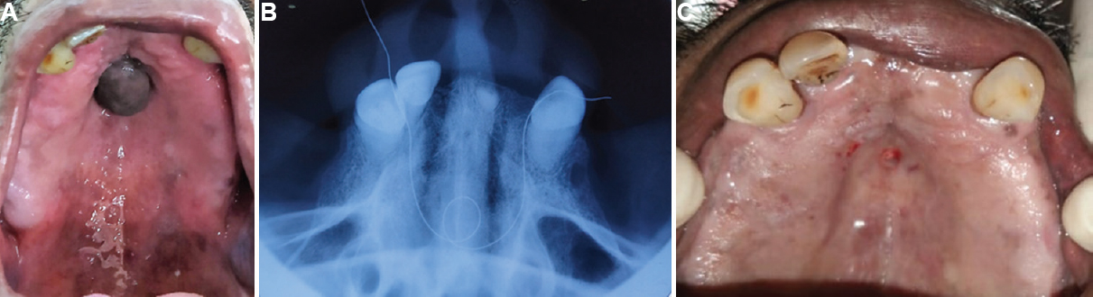
- (A) Pre-operative picture showing growth in the mid-palate. (B) Occlusal radiograph with string showing no bony involvement. (C) Picture showing immediately after excision of growth in the mid-palate.
Clinically, the palatal growth was diagnosed as fibroma. Maxillary occlusal radiograph with string around the growth was taken which revealed no bony involvement (Fig. 1B). The patient's medical history was non-contributory, and blood investigations were within normal limits. Excisional biopsy of the palatal growth was planned, and excision of the growth was done under local anaesthesia (Fig. 1C). Histopathological examination of the excised palatal growth (Fig. 2A and B) confirmed the clinical diagnosis of fibroma. The patient was under regular follow up for one month. No recurrence was seen till then with satisfactory healing. The patient was advised for complete denture prosthesis after extraction of periodontally compromised teeth.
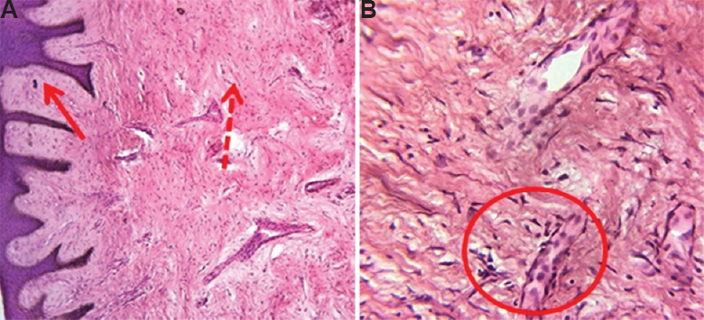
- (A) Photomicrograph showing dense bundles of collagen fibres (dotted arrow) with stratified squamous epithelium (arrow) (H and E, ×10). (B) Abundance of spindle-shaped fibroblasts was seen (inside the circle) which are characteristics of fibroma (H and E, ×40).
Acknowledgment:
Authors acknowledge the department of Oral Pathology, Tamil Nadu Government Dental College and Hospital, Chennai, Tamil Nadu, for histopathological examination of the tissue.
Conflicts of Interest: None.


