
Translate this page into:
Osteopetrosis with mandibular osteomyelitis
* For correspondence: drsjayachandranmds@yahoo.com
-
Received: ,
This article was originally published by Wolters Kluwer - Medknow and was migrated to Scientific Scholar after the change of Publisher.
A three year old female† child was brought to the department of Oral Medicine and Radiology, Tamil Nadu Government Dental College and Hospital, Chennai, India, in September 2019, with chief complaints of pain and purulent discharge from the jaw. General examination revealed squint, nystagmus, wide nasal bridge, macrocephaly, splenomegaly and hepatomegaly. Local examination showed sinus opening in the left side of the mandible with purulent discharge (Fig. 1A), presence of extensive carious tooth with exposed root in the maxilla and exposed alveolar bone with denuded mucosa in the mandible (Fig. 1B). She was anaemic with low haemoglobin of 4.2 gm/dl and raised erythrocyte sedimentation rate of 16 mm/h. Panoramic radiograph revealed increased bone density, unerupted tooth buds, areas of radiolucencies with bone resorption, suggestive of osteomyelitic changes (Fig. 2A). Thickened calvarium (Fig. 2B), bone within the bone appearance of ribs (Fig. 2C), increased density of bones with reduction of bone marrow (Fig. 2D), sandwich appearance of vertebrae and Erlenmeyer flask deformity of the femur were seen on skeletal survey supporting the diagnosis of infantile osteopetrosis with secondary osteomyelitis of the mandible. Local debridement using diluted hydrogen peroxide 20 per cent volume was done, and meticulous oral hygiene measures were advised. Following interdisciplinary consultation with the department of Pediatrics, Institute of Child Health and Hospital for children, Chennai, she was treated with intravenous injection of ceftriaxone 375 mg twice daily, injection of vancomycin 200 mg thrice daily, syrup paracetamol 5 ml (only when the patient has pain) for six weeks. After six weeks of antibiotic treatment, improvement in the healing of sinus was observed (Fig. 3). Thereafter, the patient was followed up for six months periodically for about two years by teleconsultation; the patient is healthy without any recurrences during the period of follow up.
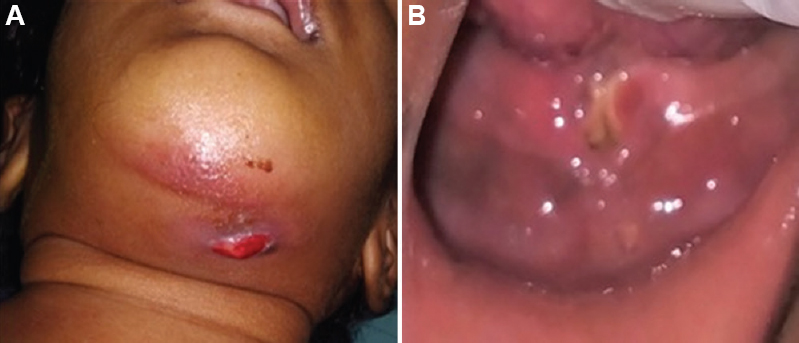
- (A) Image showing draining sinus in the left side of the mandible. (B) Image showing exposed alveolar bone in the mandible with denuded mucosa.
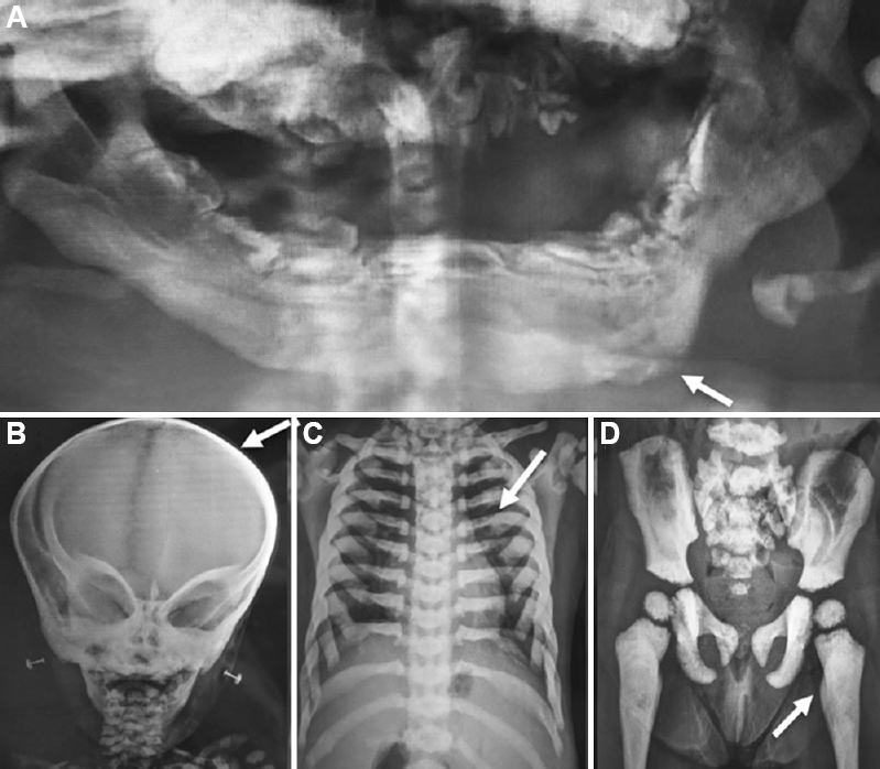
- (A) Panoramic radiograph showing maxilla and mandible alveolar bone with multiple unerupted malformed tooth buds and mixed radiolucency and radiopacity with resorption of the lower border of the left side of the mandible (arrow). (B) Image showing increased thickening of the calvarium (arrow). (C) Image showing bone within the bone appearance of ribs. (D) Image showing the increased density of bones with reduced bone marrow (arrow).
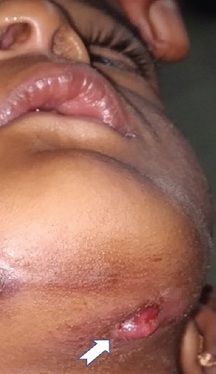
- Image showing the healing sinus in the left side of the mandible.
Acknowledgment
Authors acknowledge Dr Sibhisrivathsan, Institute of Child Health, Egmore, Chennai, for the clinical management of the patient.
Financial support & sponsorship: None.
Conflicts of Interest: None.


