
Translate this page into:
Orofacial granulomatosis - Intralesional steroid therapy
*For correspondence: dr.kotyanaik.maloth@gmail.com
-
Received: ,
This is an open access journal, and articles are distributed under the terms of the Creative Commons Attribution-NonCommercial-ShareAlike 4.0 License, which allows others to remix, tweak, and build upon the work non-commercially, as long as appropriate credit is given and the new creations are licensed under the identical terms.
This article was originally published by Wolters Kluwer - Medknow and was migrated to Scientific Scholar after the change of Publisher.
A 55 yr old female† presented to the department of Oral Medicine and Radiology, Mamata Dental College, Khammam, Telangana, India, in June 2019, with the complaints of progressive swelling on her lower and middle third part of the face for the last four years. No history of food allergy was recorded.
On extra-oral examination, a diffuse swelling was seen on her lower and middle third of the face, involving upper lip, lower lip, chin and cheek region. Skin over the swelling was slightly erythematous with shiny surface, firm and non-tender (Fig. 1A). On intra-oral examination, generalized oedematous and erythematous gingiva was noted. Swelling involving the labial mucosa of both upper and lower lips and right and left buccal mucosa was seen. On palpation, it was firm and non-tender (Fig. 1B). Baseline investigations were non-contributory. Incisional biopsy was done with respect to upper lip. Haematoxylin and eosin-stained histopathology pictures revealed that superficial lamina propria consists of areas of non-caseating granulomas along with dilated blood capillaries and lymphocytes, suggestive of orofacial granulomatosis (Fig. 2).
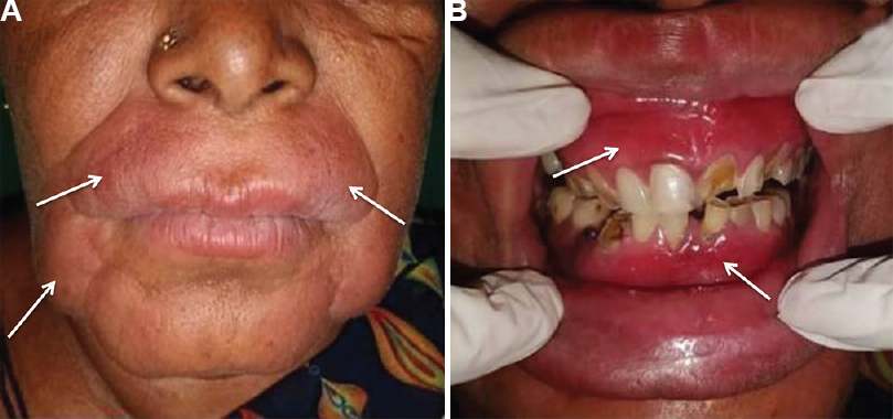
- (A) Swelling on her lower and middle third of the face (arrows). (B) generalized oedematous and erythematous gingiva with respect to both upper and lower arch (arrows).
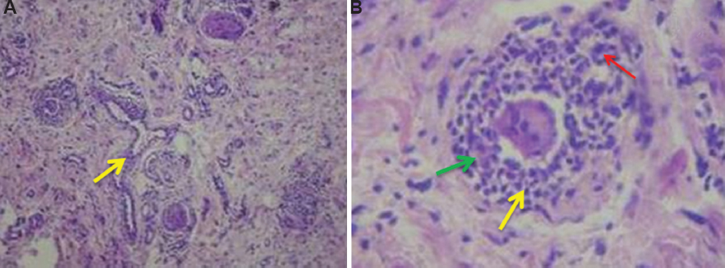
- Haematoxylin and eosin-stained histopathology pictures revealed (A) non-caseating granulomas along with dilated blood capillaries (yellow arrow) and lymphocytes (×10). (B) non-caseating granulomas are surrounded by histocytes (green arrow), lymphocytes (yellow arrow), blood vessels and multinucleated giant cells (red arrow) (×40).
A combination of intralesional injection of corticosteroids [injection triamcinolone acetonide (Kenocort)] 10 mg/ml twice a week for three weeks was started, along with tablet minocycline (OD) 100 mg for three weeks. Post-treatment follow up after three weeks showed complete resolution of swelling without recurrence (Fig. 3).
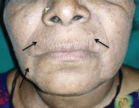
- Post-treatment follow up at the end of third week (arrows).
Conflicts of Interest: None.


