
Translate this page into:
Nasal mass in pregnancy
*For correspondence: asmitamadhavi@yahoo.co.in
-
Received: ,
This is an open access journal, and articles are distributed under the terms of the Creative Commons Attribution-NonCommercial-ShareAlike 4.0 License, which allows others to remix, tweak, and build upon the work non-commercially, as long as appropriate credit is given and the new creations are licensed under the identical terms.
This article was originally published by Wolters Kluwer - Medknow and was migrated to Scientific Scholar after the change of Publisher.
A 20 yr old female† at 37 wk of gestation presented to the department of ENT, King Edward Memorial Hospital, Mumbai, India, in October 2019, with a left-sided bleeding nasal mass. Initially, it was small in size but aggressively increased over a fortnight (Fig. 1). Magnetic resonance imaging was done to avoid radiation exposure which was suggestive of a soft tissue lesion antero-inferior to nasal septum (Fig. 2). Complete excision was done with cauterization of the base (Fig. 3), endoscopically (Fig. 4 and Video). Histopathological report was suggestive of haemangioma. Nasal mucosa on follow up after five days healed well.
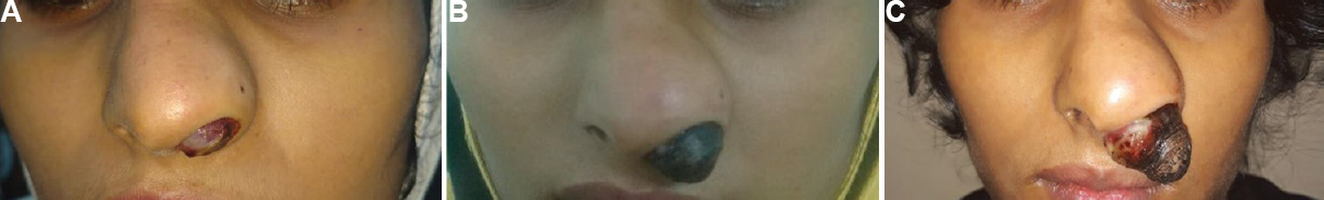
- (A) Widening of vestibule with firm nasal mass protruding from the left nostril covered with mucoid discharge on day 3 of presentation.(B) Nasal mass with externally exposed part showing necrosis and crusting on day 9 of presentation. (C) Increase in size of mass with crusting and necrosis on day 14 of presentation.
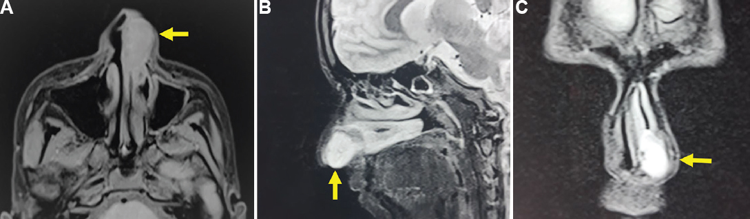
- (A) Magnetic resonance imaging – a 24×11×21mm, isointense homogenous, well-defined mass in the left nasal cavity abutting the inferior turbinate posteriorly. (B) Magnetic resonance imaging – Nasal mass arising anteroinferior to septal cartilage. (C) Magnetic resonance imaging – Hyperintense nasal mass confined to the vestibule of the left nasal cavity.
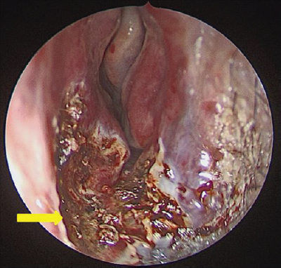
- Intraoperative cavity after complete endoscopic excision of nasal mass. Arrow showing site of attachment.
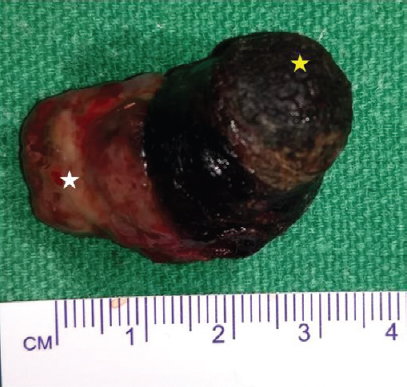
- Specimen of excised nasal mass showing intranasal vascular mucosa (white star) and external necrosed part of mass (yellow star).
Pyogenic granuloma and haemangiomas are the most common benign vascular tumours in pregnancy. Nasal mass during pregnancy has aggressive growth under hormonal influence. These are notorious as these progresses rapidly over a short duration under the influence of oestrogen as a mediator for vascular proliferation. Hence, it requires aggressive management to avoid further complications due to anaemia.
 Video available at ijmr.org.in.
Conflicts of Interest: None.


