
Translate this page into:
Metastatic calcification in end-stage renal disease: Contra naturam
*For correspondence: inderpgi@outlook.com
-
Received: ,
This is an open access journal, and articles are distributed under the terms of the Creative Commons Attribution-NonCommercial-ShareAlike 4.0 License, which allows others to remix, tweak, and build upon the work non-commercially, as long as appropriate credit is given and the new creations are licensed under the identical terms.
This article was originally published by Wolters Kluwer - Medknow and was migrated to Scientific Scholar after the change of Publisher.
A 16 yr old male child† with end-stage renal disease on maintenance haemodialysis presented in January 2019, with a history of dyspnoea and dry cough of three-month duration to the Chest Clinic of Postgraduate Institute of Medical Education & Research, Chandigarh, India.
His serum creatine was 8 mg/dl. His serum calcium and phosphorus was 11.2 mg/dl and 5.6 mg/dl, respectively. His serum parathyroid hormone level was 500 pg/ml. A computed tomography of the thorax revealed bilateral ground-glass opacification in the lung and calcification of the subcutaneous vessels (Fig. 1A and B). X-ray of the upper and lower limbs also revealed calcification of the vessels (Fig. 1C and D). A transbronchial lung biopsy demonstrated diffuse calcification of the lung interstitium and the vessel wall (Fig. 2), confirming a diagnosis of metastatic calcification. He underwent para-thyroidectomy and was referred for renal transplantation. Pulmonary calcification is rare in end-stage renal disease. Knowledge of the radiological findings of metastatic pulmonary calcification can help in avoiding invasive procedures such as bronchoscopy.
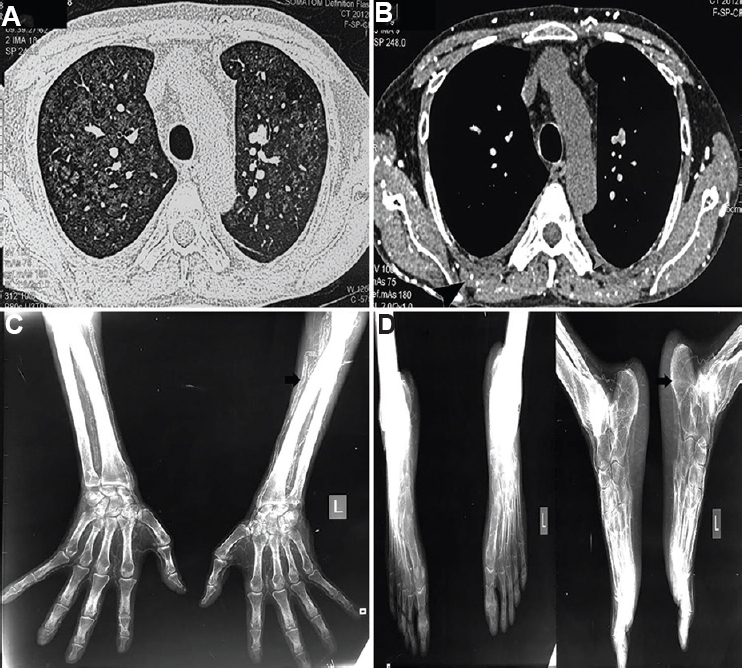
- High-resolution computed tomography of the thorax demonstrating (A) diffuse ground-glass opacification in the lung window and (B) multiple calcified vessels (arrowhead) in the mediastinal window. X-ray of the bilateral (C) upper and (D) lower limbs revealing arterial calcification of arteries (arrows).
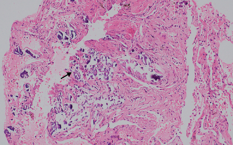
- Haematoxylin and eosin (×200) stain of the transbronchial lung biopsy demonstrating calcification (arrow) in the interstitium and the vessel wall, confirming it to be a case of metastatic calcification.
Conflicts of Interest: None.


