
Translate this page into:
Massive cerebral infarct due to Trousseau's syndrome in gastric cancer
*For correspondence: drcamans@gmail.com
-
Received: ,
This is an open access article distributed under the terms of the Creative Commons Attribution-NonCommercial-ShareAlike 3.0 License, which allows others to remix, tweak, and build upon the work non-commercially, as long as the author is credited and the new creations are licensed under the identical terms.
This article was originally published by Medknow Publications & Media Pvt Ltd and was migrated to Scientific Scholar after the change of Publisher.
A 27 yr old woman presented to the department of Medicine, MES Medical College, Perinthalmanna, Kerala, India, in January 2015 with sudden onset left-sided hemiplegia in the morning. She had advanced signet ring cell carcinoma of the stomach diagnosed 20 days earlier and received one cycle of chemotherapy. Her routine blood investigations were normal. Magnetic resonance imaging of brain revealed large infarct in right middle cerebral artery territory (Figs 1 & 2). Tests for antinuclear antibody and antiphospholipid antibodies were negative. Hereditary hypercoagulable states including protein C, protein S, antithrombin III deficiencies and activated protein C resistance were negative. Echocardiogram and carotid vertebral Doppler were normal. A diagnosis of cerebral infarct due to Trousseau's syndrome was made since other causes were ruled out. She was started on anticoagulants. Her hemiplegia persisted when followed up after two months. Trousseau's syndrome involves unexplained thrombotic events preceding the diagnosis of an occult visceral malignancy or with the tumour. Gastric malignancy leading to Trousseau's syndrome is rare.
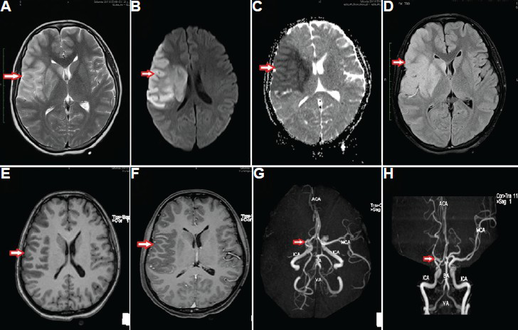
- Contrast magnetic resonance imaging of brain: Large area of T2W and fluid-attenuated inversion recovery hyperintensity (arrow in A and B) in the right fronto-temporo-parietal region with diffusion restriction (arrow in C) appearing hypointense in apparent diffusion coefficient (arrow in D) without contrast enhancement (arrows in E and F). Magnetic resonance angiogram showed absent signal in the right middle cerebral artery (arrows in G and H).
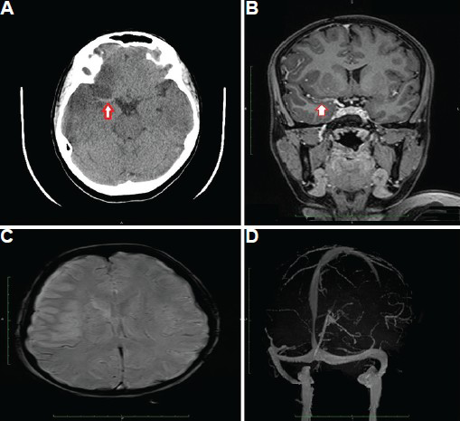
- Contrast magnetic resonance imaging of brain: Axial computed tomography scan image (A) and coronal T1 fluid-attenuated inversion recovery image (B) showing thrombus in M1 segment of the right middle cerebral artery (arrows). Susceptibility weighted image (C) did not show areas of blooming and magnetic resonance venogram (D) was also normal.


