
Translate this page into:
Klebsiella pneumoniae-associated pneumorrhachis
* For correspondence: 814288678@qq.com
-
Received: ,
This article was originally published by Wolters Kluwer - Medknow and was migrated to Scientific Scholar after the change of Publisher.
A 69 yr old female† who had poorly controlled type 2 diabetes for 10 years was presented to the emergency department at Fujian Provincial Hospital, Fuzhou, PR China, in December 2020, with coma for two hours. On admission, physical examination showed that she had no fever, but blood pressure dropped to 58/36 mmHg and heart rate increased to 157 beats per min. Blood tests showed an increase in the levels of leucocyte count, procalcitonin, glucose and lactic acid to 13.0×10⁹/l, 27.0 μg/l, 34.0 mmol/l and 6.0 mmol/l, respectively. Laboratory tests also indicated that the patient displayed liver, renal, cardiac and respiratory dysfunction. Emergency computed tomography (CT) was performed which showed pneumorrhachis, concurrent with emphysematous diaphragmitis and psoas infection (Figs 1 and 2). She was diagnosed with septic shock and multiple organ dysfunction syndrome (MODS) and immediately received empirical anti-infection [imipenem and cilastatin sodium, (IMP/CS)], glycaemic control, anti-shock and organ support treatments. Klebsiella pneumoniae susceptible to IMP/CS was detected from the blood by bacterial culture. However, the patient’s condition deteriorated rapidly and she died of septic shock and MODS one day after admission.
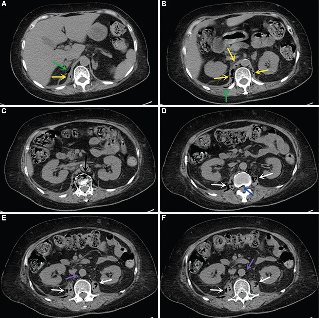
- Axial CT images showing pneumatosis in diaphragm (Panels A and B, yellow arrows) and surrounding space (Panels A and B, green arrows), vertebra and paravertebral space (Panel C, black arrow), intraspinal canal (Panel D, blue arrow), bilateral psoas (Panels D-F, white arrows) and para-aortic space (Panels E and F, purple arrows). CT, computed tomography.
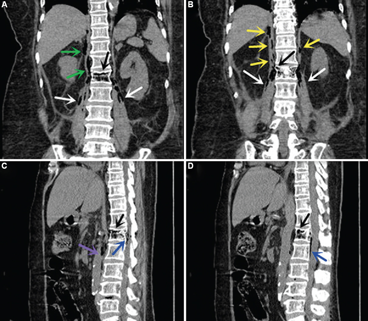
- Coronal and sagittal CT images using multi-planar reformatting showing emphysematous infection in the diaphragm (Panel B, yellow arrows) and surrounding space (Panel A, green arrows), vertebra (Panels A-D, black arrows), intraspinal canal (Panels C and D, blue arrows), bilateral psoas (Panels A and B, white arrows) and para-aortic space (Panel C, purple arrow). CT, computed tomography.
K. pneumoniae is a kind of fermentative bacterium and high levels of blood glucose provide it with favourable conditions for haematogenous dissemination, colonization, growth and aerogenesis in local tissues, which eventually leads to tissue destruction, endotoxaemia, septic shock and MODS. Pneumorrhachis may indicate fermentative bacterial infection in patients who present with hyperglycaemia, septic shock and MODS, which would be helpful for guiding empirical anti-infection treatment before pathogenic bacteria are isolated.
Financial support and sponsorship
None.
Conflicts of interest
None.
Acknowledgment:
Authors acknowledge Dr Ruiping Cai, department of Radiology, Fujian Provincial Hospital, Fuzhou, PR China, for providing the CT images.


