
Translate this page into:
Intraoral malignant melanoma
* For correspondence: dr.neelammittal@gmail.com
-
Received: ,
This is an open access article distributed under the terms of the Creative Commons Attribution-NonCommercial-ShareAlike 3.0 License, which allows others to remix, tweak, and build upon the work non-commercially, as long as the author is credited and the new creations are licensed under the identical terms.
This article was originally published by Medknow Publications & Media Pvt Ltd and was migrated to Scientific Scholar after the change of Publisher.
A 26 yr old female presented to the Faculty of Dental Sciences, Institute of Medical Sciences, Banaras Hindu University, Varanasi, India, in September 2015, with black discolouration of her upper gums both on labial and palatal sides. According to the patient this was present for the past four months. There was no history of smoking and drinking. A diffuse black pigmentation on the labial and palatal aspects of maxillary arch on its anterior part (Figs 1 & 2) was seen. There was no tooth mobility on examination and also lymph nodes were not palpable. The condition was initially suspected to be physiological pigmentation, but to make confirmatory diagnosis, incisional biopsy was done that confirmed the lesion to be malignant melanoma (Fig. 3). Primary malignant melanoma is less common in the oral cavity and is usually asymptomatic in early stages that make its diagnosis difficult. The patient was referred to a higher centre for further management.
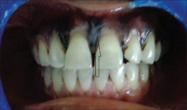
- Black pigmentation on the labial aspect of maxilla (arrow).
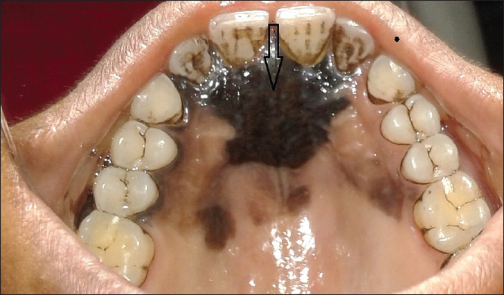
- Black pigmentation on the palatal aspect of maxilla (arrow).
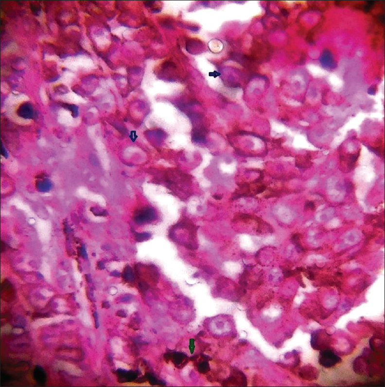
- Photomicrograph of hematoxylin and eosin (H&E) stained section (×100) showing haphazardly distributed atypical melanocytes having high N/C (nucleus/cytoplasm) ratio with macronuclei (blue arrows) and melanin pigment secreted by melanocytes (green arrow).


