
Translate this page into:
Giant virilizing adrenocarcinoma: Rare presentation & management dilemma
* For correspondence: smruti63@hotmail.com
This is an open access article distributed under the terms of the Creative Commons Attribution-NonCommercial-ShareAlike 3.0 License, which allows others to remix, tweak, and build upon the work non-commercially, as long as the author is credited and the new creations are licensed under the identical terms.
This article was originally published by Medknow Publications & Media Pvt Ltd and was migrated to Scientific Scholar after the change of Publisher.
A 40 year old female, first presented in August 2012 in Topiwala National Medical College (TNMC) and BYL Nair Hospital, Mumbai, India, in surgical outpatients, with a left flank lump, oligomenorrhoea, weight loss, increased facial hair and deepening of voice for the last two months. The abdomen was distended, soft, non-tender, with a firm ballotable 17x10 cm mass moving with respiration palpable in left flank. Computed tomography (CT) abdomen was suggestive of 17x13x12 cm well defined heterogenously enhancing mass in left suprarenal region. Fat planes with left kidney, spleen and tail of pancreas were lost (Fig. 1).
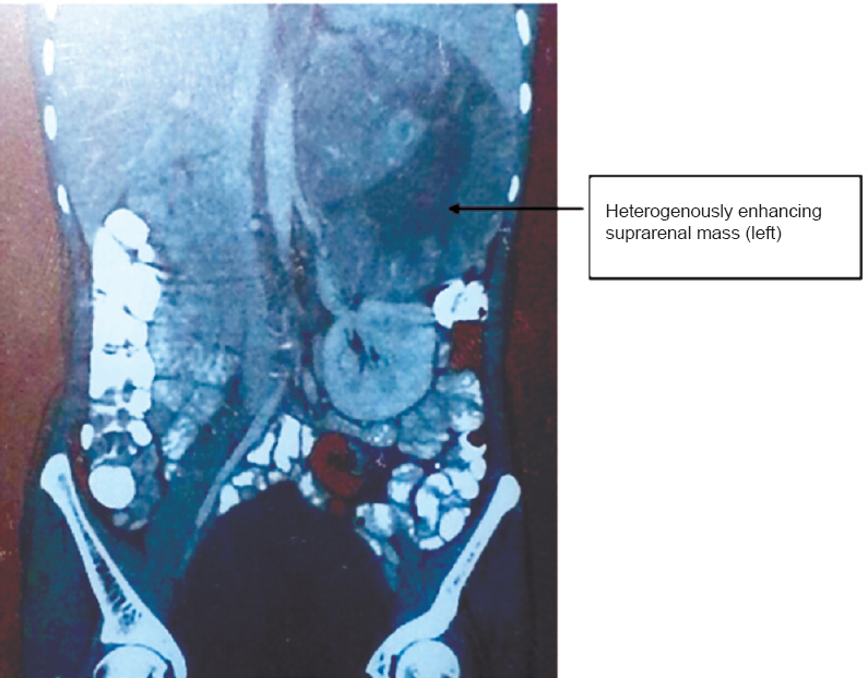
- CT scan showing large well defined heterogeneously enhancing mass in left suprarenal region with loss of fat planes between the mass, spleen and left kidney.
Baseline investigations were normal, ruling out a cortisol secreting adrenocortical tumour or a pheochromocytoma. The virilising features were attributed to raised levels of dehydroepiandrosterone sulphate [DHEAS, >1000 pg/dl (ref. range 32-240)].
The diagnosis of a functional adrenal tumour was made and decision was taken to remove the tumour as it was still resectable. As tumour mass was large, transperitoneal approach was taken. Approximately 20x12x10 cm left suprarenal mass adhered to the left kidney and spleen was found, pancreas was not involved. Adrenal mass with left kidney and spleen was removed (Fig. 2). Postoperative mitotane based chemotherapy was given.
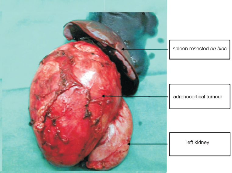
- Post-operative specimen showing the adrenal tumour attached to left kidney and spleen.
Follow up CT abdomen at six months and one year was not suggestive of any recurrence. The patient showed gradual, steady and complete improvement from the virilizing features. DHEAS levels performed postoperatively at six months and one year interval were normal. This was a rare case where a giant adrenocarcinoma was surgically resected with curative intent without neoadjuvant chemotherapy.


