
Translate this page into:
Freely moving right ventricular thrombus in a patient with acute pulmonary embolism
*For correspondence: negiprakash59@gmail.com
-
Received: ,
This is an open access article distributed under the terms of the Creative Commons Attribution-NonCommercial-ShareAlike 3.0 License, which allows others to remix, tweak, and build upon the work non-commercially, as long as the author is credited and the new creations are licensed under the identical terms.
This article was originally published by Medknow Publications & Media Pvt Ltd and was migrated to Scientific Scholar after the change of Publisher.
A 28 yr old female patient presented to the department of Cardiology, Indira Gandhi Medical College, Shimla, India, in December 2015, with complaints of sudden onset dyspnoea of two days duration. There was no chest pain, cough or fever. Patient had been using oral contraceptives for the past five months. She was having tachycardia (114 bpm) and a blood pressure of 126/80 mmHg. Chest and cardiovascular examination was unremarkable. Electrocardiogram revealed sinus tachycardia. Serum D-dimer was elevated. She was subjected to computerized pulmonary angiography which showed a thrombus in right pulmonary artery (Figure A, arrow). Echocardiography showed normal right ventricular function but demonstrated a highly mobile-free floating thrombus in the right ventricular cavity (Figure B and Video). Cardiac magnetic resonance imaging (MRI) confirmed it to be a free-floating thrombus and ruled out tumour (Figure C). Duplex scan of lower limbs ruled out deep venous thrombosis. Since patient was haemodynamically stable and had normal right ventricle function, she was treated with low molecular weight heparin and warfarin. There was complete dissolution of thrombus in three week time (Figure D). Detailed workup for the genetic and acquired causes of thrombophilia was negative except for the history of oral contraceptive use. Careful evaluation is warranted for better identification.
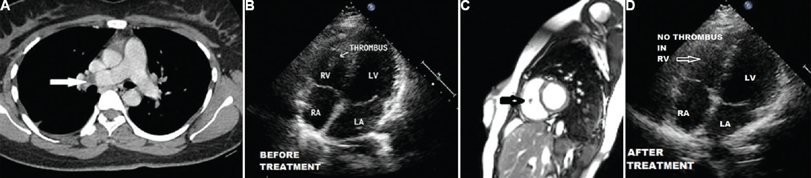
- (A) Computed tomography pulmonary angiogram showing a filling defect (arrow) in the distal right pulmonary artery. (B) Transthoracic echocardiogram showing a thrombus (arrow) in the right ventricle. (C) Cardiac magnetic resonance image showing a stalk-free mass suggestive of thrombus in the right ventricle (arrow). (D) Transthoracic echocardiogram, three-week post-treatment, showing complete resolution of the right ventricular thrombus. RV & LV, right and left ventricle; RA & LA, right and left atrium.


