
Translate this page into:
Fitz-Hugh-Curtis syndrome
*For correspondence: rathipmpp@gmail.com
-
Received: ,
This is an open access article distributed under the terms of the Creative Commons Attribution-NonCommercial-ShareAlike 3.0 License, which allows others to remix, tweak, and build upon the work non-commercially, as long as the author is credited and the new creations are licensed under the identical terms.
This article was originally published by Medknow Publications & Media Pvt Ltd and was migrated to Scientific Scholar after the change of Publisher.
A 23 yr old woman presented to the department of Gastroenterology at Bombay Hospital, Mumbai, India, in July 2015 with an acute onset, sharp right hypochondriac pain; intermittent fever and gradually progressive symmetrical abdominal distension for the preceding two weeks. On physical examination, ascites was evident. On analysis, the ascitic fluid was culture-negative and exudative with normal adenosine deaminase value. C-reactive protein was elevated. Her blood culture and test for human immunodeficiency virus were negative. A computerized tomogram of the abdomen was performed (Fig. 1). She underwent an exploratory laparoscopy that revealed flimsy adhesions between the inter-bowel loops (Fig. 2), between the abdominal wall and bowel loops and along the liver surface, ascites and features suggestive of perihepatitis and right salpingitis. Based on these findings, the diagnosis of Fitz-Hugh-Curtis syndrome was made. An endocervical swab did not reveal any pathological organisms as the patient by then had received seven days of intravenous cephalosporins. She was discharged on oral azithromycin. On follow up after one month, she was asymptomatic.
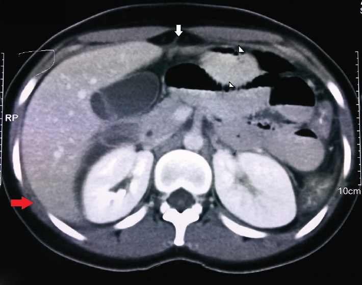
- A computerized tomogram of the abdomen revealed a thickened liver capsule and subcapsular fluid (red arrow), suspicious adhesions between the liver capsule and anterior peritoneum (vertical arrow) and between the inter-bowel loops (arrowheads), diffuse colonic wall thickening and moderate ascites.
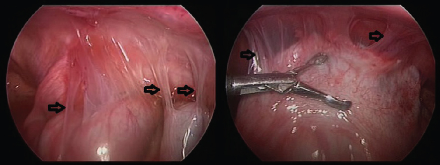
- The flimsy “violin string-like adhesions” on laparoscopic examination (arrows).


