Translate this page into:
Expression of γH2AX may help in defining a genetically more stable subtype of infiltrating ductal carcinoma of breast
Reprint requests: Dr Alka Bhatia, Assistant Professor, Department of Experimental Medicine & Biotechnology Postgraduate Institute of Medical Education & Research, Chandigarh 160 012, India e-mail: alkabhatia@ymail.com
-
Received: ,
This is an open-access article distributed under the terms of the Creative Commons Attribution-Noncommercial-Share Alike 3.0 Unported, which permits unrestricted use, distribution, and reproduction in any medium, provided the original work is properly cited.
This article was originally published by Medknow Publications & Media Pvt Ltd and was migrated to Scientific Scholar after the change of Publisher.
Abstract
Background & objectives:
Gamma H2AX, a marker of DNA double stranded breaks (DSB) has been found to be over expressed in various tumours. The objective of the present work was to study the expression of γH2AX in infiltrating ductal carcinoma (IDC) and fibroadenoma (FA) cases and to associate the expression in IDC with cytomorphological features and DNA ploidy.
Methods:
The expression of γH2AX was studied in fine needle aspirates of 16 cases of IDC and 15 FA cases. The expression in IDC was correlated with the cytological grade, apoptotic (AI) and mitotic indices (MI) and ploidy status.
Results:
A high γH2AX expression was noted in IDC as compared to FA. Amongst the IDC cases the γH2AX was found to be significantly over expressed in DNA diploid IDC cases as compared to the aneuploid ones.
Interpretation & conclusions:
The study suggests a role of γH2AX in breast carcinogenesis which needs to be explored further. Moreover, the γH2AX expression together with ploidy status may serve as a means of assigning the patients of IDC to a better prognostic category irrespective of the cytomorphogical parameters.
Keywords
Apoptosis
cytology
fibroadenoma
γH2AX
grading
IDC
ploidy

In eukaryotic cells, the role of histone proteins in organization of chromatin is well-recognized. H2AX is a histone variant belonging to H2A family which comprises 2-10 per cent of the H2A complement in the mammalian tissues1. H2AX is activated when DNA molecules inside the cell are broken by external factors such as radiation or sometimes spontaneously as in tumours2. H2AX becomes phosphorylated at serine 139 residue in chromatin surrounding DNA double-strand breaks (DSBs) to form Gamma H2AX (γH2AX)1. The phosphorylation process is rapid and extensive, covering large regions of the chromosome adjacent to each break and producing foci that can be visualized microscopically after antibody labelling31. γH2AX is proposed to concentrate repair factors at sites of DNA damage and maintain genomic stability4. Animal experiments have shown increased incidence of genomic instability and tumour formation in case of partial or complete loss of H2AX gene5. Therefore, H2AX has been proposed to act as a tumour suppressor in concert with other tumour suppressors like p53. The soluble H2AX, that is, the non-chromatin-associated H2AX has been found to sensitize cells with DNA damage to undergo apoptosis6. The DNA damage response pathway is activated early in the process of carcinogenesis even before activation of p537. Hence it has been proposed that antibody to γH2AX can be used to detect precancerous lesions or as a biomarker for cancer detection at the metastasis site89.
It is now well known that genetic factors play an important role in development of invasive breast cancer10. A sequential accumulation of genetic alterations involving activation of oncogenes and by inactivation of several tumour suppressor genes has been found in this malignancy10. More recent studies have suggested that mechanisms and pathways regulating DNA damage and repair may play a mutator role in driving breast cancer pathogenesis11. Though preliminary work has shown increased expression of γH2AX in tumours of breast, melanoma, prostate, and urinary bladder, information is limited121314. Moreover, a systematic correlation with other significant parameters like grade, apoptosis, mitosis and ploidy status has not been studied before in breast cancer. The present work was therefore, undertaken to study the expression of γH2AX in infiltrating ductal carcinoma (IDC) and fibroadenoma (FA) cases and to relate the expression in IDC with cytomorphological features and DNA ploidy.
Material & Methods
This prospective study involved 53 patients with a palpable breast lump coming to Fine Needle Aspiration Cytology (FNAC) clinic of department of Cytopathology or Surgery, Postgraduate Institute of Medical Education & Research, Chandigarh, a tertiary care hospital between May 2009 and March 2011. The work was carried out after obtaining ethical clearance by Institute's ethics review committee and after an informed written consent was obtained from all the patients.
Only fresh cases without any history of breast cancer in the family were included in the study. Those on any kind of treatment for breast disease were excluded. Fine needle aspirate was collected by the standard protocol of breast FNAC and was utilized for making the cytology smears and for performing flow cytometry. The smears were stained with Giemsa for cytological diagnosis and grading as per the grading system proposed by Robinson et al15. Apoptotic and mitotic indices (AI & MI) were determined by counting 1000 tumour cells and calculating the percentage in each case of IDC. For studying the γH2AX expression, cell suspension of the aspirate was prepared in phosphate buffered saline (PBS) pH 7.4. The cell concentration (2×10⁶) was checked by counting the number of cells using a haemocytometer. After fixation in 1 per cent formaldehyde- PBS (pH 7.4) and permeabilization in cold ethanol, immunostaining was carried out by incubating the cells with 200 μl mouse monoclonal FITC conjugated anti-phosphohistone γH2AX antibody (1:500 dilution, Biolegend, San Diego, USA). The stained cells were then analysed under flow cytometer (BD FACS Calibur, 2002, USA) using BD cell quest™ pro data software. The instrument was calibrated using Calibrite beads. Approximately 10,000 events were collected from each sample for analysis. The ploidy status and S-phase fraction (SPF) was determined by flow cytometry after incubation with propidium iodide (PI). Statistical analysis of the data was carried out using SPSS programme (version 16) IBM, USA using independent t test, ANOVA test and Pearson's correlation analysis.
Results
Out of 53 cases included in the study, a cytological diagnosis of IDC was offered in 21 cases and 17 cases were reported as fibroadenoma (FA). The age range of patients was 19-76 yr, median age being 38 yr. The age was between 35-76 yr (median 40.5 yr) for IDC and 19-65 yr (median 28 yr) for FA. The lump size ranged from 0.5-10 cm with a mean of 3.4 cm. The mean lump size was greater (4.02 cm) for malignancy than in FA (2.58 cm).
γH2AX Expression in FA and IDC: γH2AX expression was analysed in 16 cases of IDC and 15 cases of FA. The mean and median γH2AX expression was found to be higher in IDC (28.30 and 20.35%, respectively) and lower in FA (17.07 & 6.68%, respectively) (Fig. 1). However, the difference in γH2AX expression in malignant and benign cases was not significant.
Morphological parameters in IDC
Apoptotic and mitotic index: The apoptosis was counted morphologically in the Giemsa stained smears in 21 cases of IDC. The AI was expressed as percentage of apoptotic cells. The apoptotic cells were identified by cellular shrinkage, chromatin condensation and formation of apoptotic bodies. The AI was variable in IDC with percentage of apoptotic cells varying from 0.5 to 35 with a median of 4.5 (Table). The mitotic figures were also counted and expressed as percentage as in case of AI. The MI ranged from 0-8 with a median of 2.65 (Fig. 1).
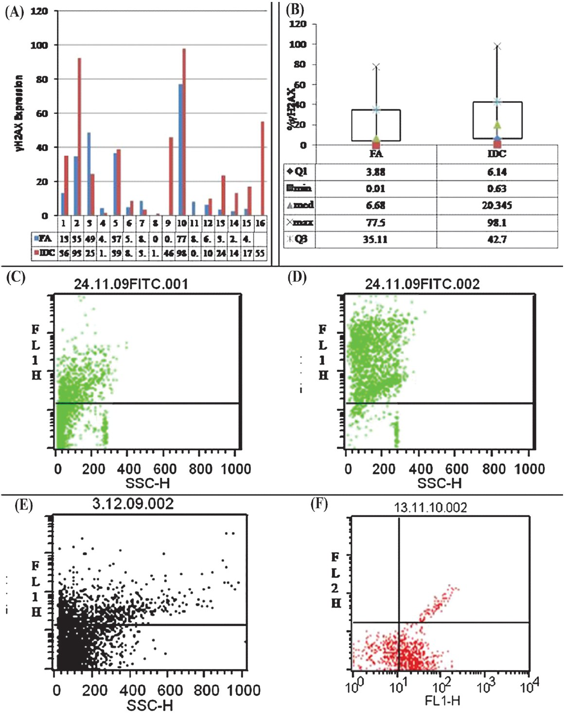
-
(A) Percentage γH2AX expression in fibroadenoma (FA) and infiltrating ductal carcinoma (IDC). (B) Box plot diagram showing the difference in median, minimum and maximum values of % γH2AX expression in FA & IDC. (C, D) % γH2AX expression (35.52 & 92.67%) in two cases of IDC seen as FITC labelled population in FL1-H channel. (E, F) Low and high % γH2AX expression (13.41 and 77.15%) in 2 cases of FA seen as FITC labelled population in FL1-H channel.
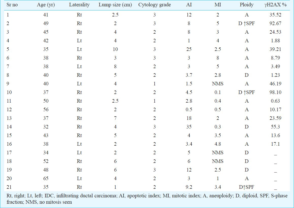
Cytological grading of IDC cases: Cytological grading was performed on all 21 fresh cases of IDC. The lesions with small cells exhibiting slight pleomorphism and many gland-like structures were classified as grade 1, those with moderate atypia and pleomorphism and 10-75 per cent gland formation as grade 2, whereas those with large cell size, marked pleomorphism, prominent nucleoli and minimal gland formation were categorized as grade 3 (Fig. 2A-C). There were two cases of grade 1, 13 cases of grade 2 and 6 cases of grade 3 IDC (Table).
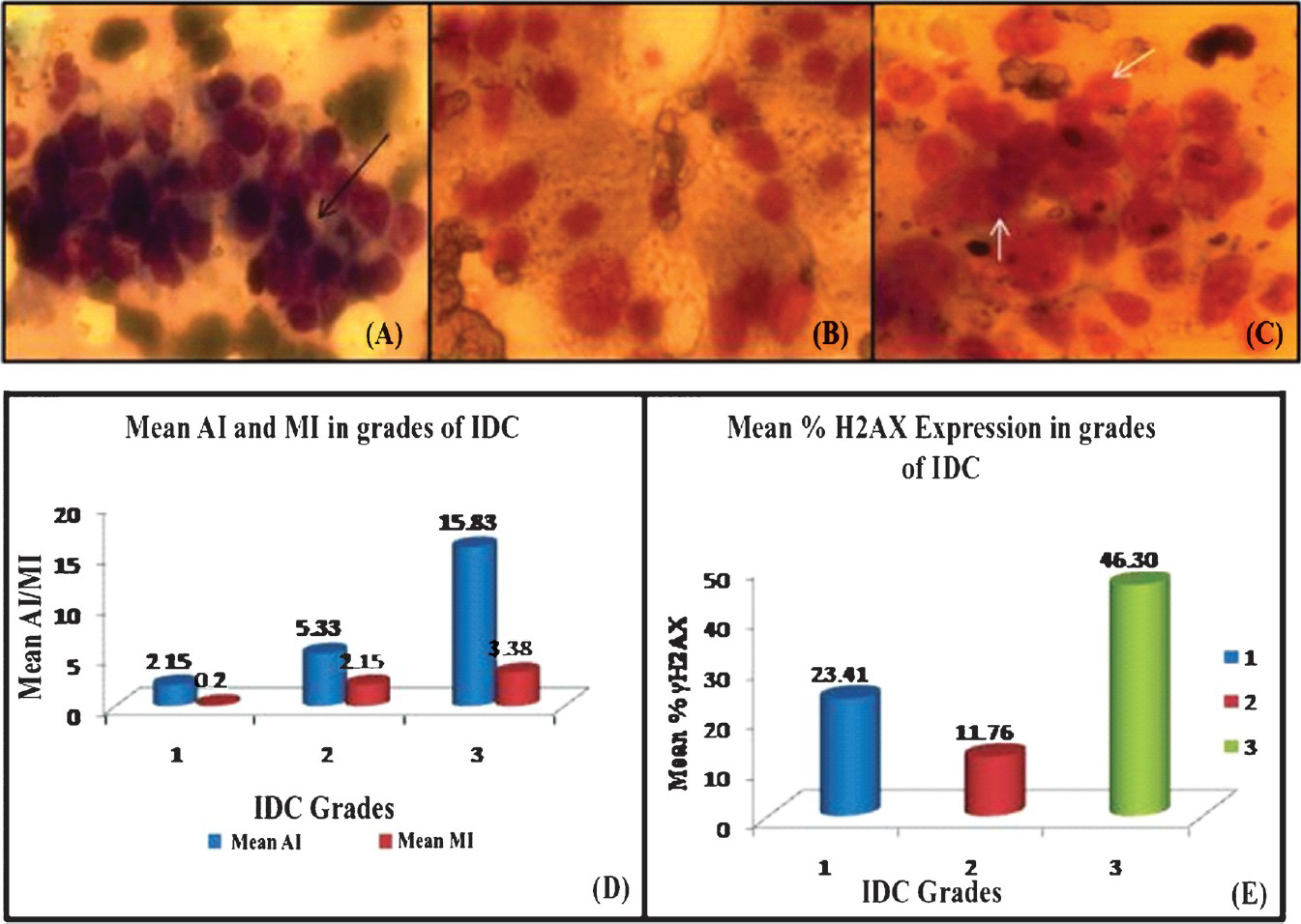
-
(A, B, C) Microphotographs showing cytological grading of IDC in FNAC smears. 2A shows grade 1 cancer with, cohesive cells, minimal pleomorphism and gland like structures (arrow black); 2B shows grade 2 cancer with moderate degree of pleomorphism and cellular discohesion; 2C shows grade 3 cancer with poorly differentiated cells exhibiting marked nuclear pleomorphism and nucleolar prominence (arrow white) (Giemsa staining) (D) Increased mean values of both AI and MI with increase in cytological grade of cancer (E) %γH2AX expression in different grades of IDC. Note the much higher expression in grade 3 as compared to the other grades of IDC.
Ploidy status in IDC: The ploidy status was analysed in 21 cases of IDC. The DNA histogram with an additional G0-G1 peak and a DNA content of <2N or >2N were classified as aneuploid and an SPF (S-phase fraction) of >10 per cent was considered to be high. In all, 13 (61.90%) cases showed aneuploidy, eight hyperdiploid aneuploidy, four hypodiploid aneuploidy and one case showed both hyper- as well as hypodiploid peak. Eight cases (38.1%) were diploid, of whom three cases showed raised SPF as well.
Correlation of cytological grades with AI and MI: The mean values of both AI and MI showed an increase with increasing grades of the IDC (Fig. 2D). On statistical analysis, the difference in AI between grade 3 and the remaining two grades was found to be significant (P<0.05). The difference in the MI of the three groups was not significant. Correlation of AI with MI in IDC cases also showed a proportional increase in both the parameters which was statistically significant (Pearson's correlation, r= .009, P<0.05).
Correlation of γH2AX expression with grade, AI, MI and ploidy status in IDC: Gamma H2AX expression in different grades of IDC showed a considerable overlap amongst individual cases belonging to each grade (Table). Mean γH2AX was found to be highest in grade 3 (46.30%) as compared to the lower grades (11.76% in grade 2 and 23.41% in grade 1, respectively) (Fig. 2E). The difference between the three groups was not significant. No significant correlation was observed between the MI and AI and γH2AX expression.
Correlation of ploidy status with γH2AX expression in IDC showed a higher expression in diploid IDC (n=4, Mean = 61.83%) especially in those with diploidy and high SPF (n=2, 92.67 & 98.10 respectively) as compared to the aneuploid cases (n = 12, Mean = 18.73%). Statistical analysis using independent t-test also showed the difference in mean values for aneuploid and diploid cases to be significant (P<0.05) (Fig. 3) The mean AI in DNA diploid cases was 12.8 as against a value of 6.9 for the aneuploid ones.
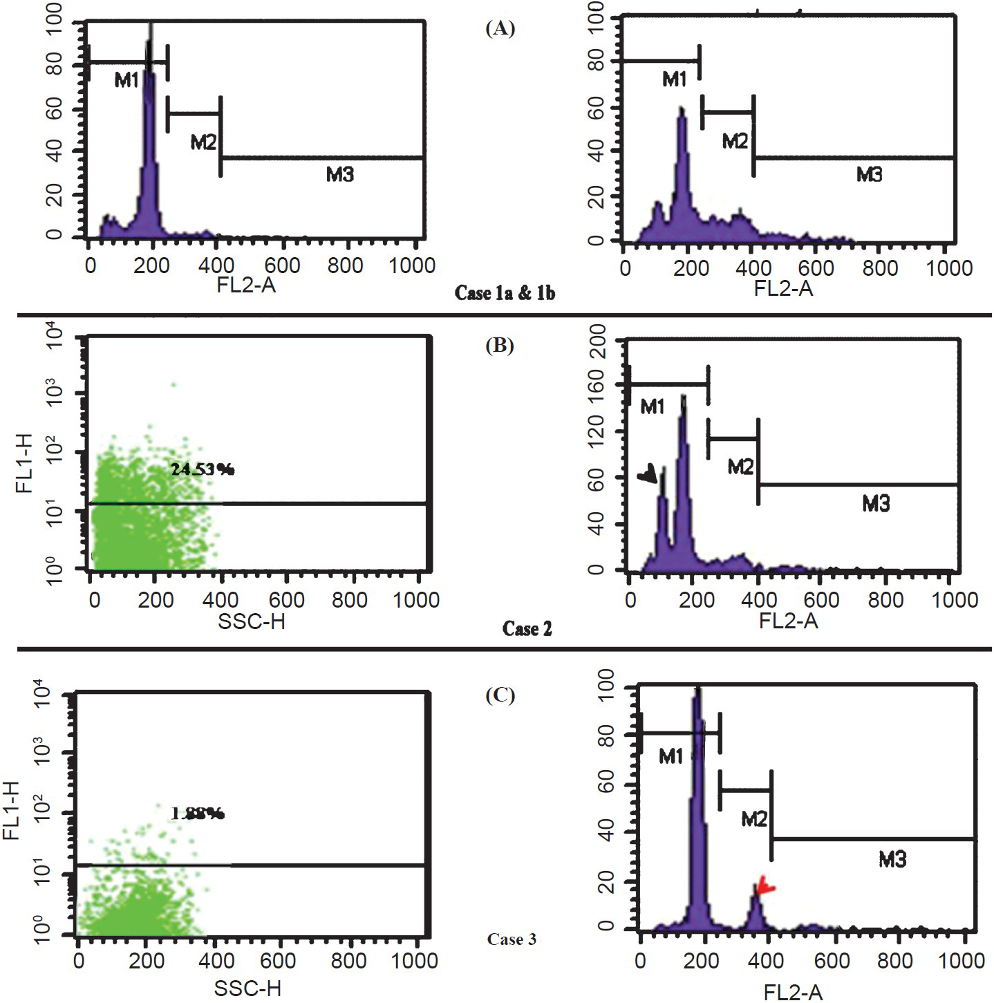
-
(A) Case 1a: DNA diploid histogram of case of FA. Corresponding γH2AX expression is shown in Fig. 1E; Case 1b: DNA diploidy with high SPF in DNA histogram of a case of IDC. Corresponding high γH2AX expression is shown in Fig. 1D (B) Case 2: A case of IDC showing hypodiploid aneuploidy (black arrowhead) with 24.53% population showing γH2AX expression (C) Case 3: A case of IDC showing small aneuploid peak and low (1.88%) γH2AX expression (red arrowhead).
Discussion
The DNA DSB is a serious lesion that can initiate chromosomal instability (CIN), ultimately leading to cancer16. DNA DSBs caused by exposure to DNA damaging agents initiate phosphorylation of histone H2AX to form γH2AX, which is considered a surrogate marker of DSBs17. In addition to being a cause of cancer, DSB induction is paradoxically an effective treatment for cancer. Many therapeutic agents act by introducing sufficient DSBs into cancer cells to activate the cell death pathways. Therefore, γH2AX expression is generally used to assess the response to chemoradiotherapy18.
In the present study, FNAC samples from IDC (untreated) and FA patients were analysed for the presence of spontaneous γH2AX foci. The mean for the two groups showed a higher value for IDC although statistically insignificant. Similar results were observed in the study by Wasco et al19 who compared the difference in expression of γH2AX in nevi and melanoma cases. This may be due to the complex mechanism by which DSBs and their repair occurs. Moreover, the detection of γH2AX foci by the antibody requires conglomeration of several foci at the break site in the absence of which a focus may not be detected20. Cheung et al14 in their study on 60 cases of low grade urothelial carcinoma found that γH2AX positivity was associated with a lower rate of tumour recurrence. However, in this study cases of recurrent cancer have not been included. Wasco and Pu21 in their study on metastatic renal cell carcinoma (RCC) concluded that γH2AX in conjunction with other antibodies may be used to detect RCC metastasis, however, in their study also a patchy staining of normal tubules was observed with γH2AX antibody21.
The overexpression of γH2AX in IDC as opposed to FA indicates its possible role in breast carcinogenesis. A previous report also confirms the presence of copy number alterations of H2AFX gene in sporadic breast cancers22. However, in view of presence of overlap amongst the FA and IDC cases in our study, the potential clinical utility of detection of γH2AX remains uncertain. Perhaps a larger series may help to highlight better the intricacies of γH2AX expression in benign and malignant lesions of the breast.
DNA ploidy has been considered as an important factor for estimating the degree of CIN which is supposed to be reflected in the form of tumour aggressiveness. Our results showed an increased expression of γH2AX in diploid IDC, more so, in diploidy with high SPF. Hau et al23 showed that polyploidy was associated with increased expression of γH2AX as compared to the diploidy. However, their study mainly included tetraploid cases rather than aneuploid ones23. Although many studies have analysed the significance of ploidy status in breast cancer, there is still controversy regarding its exact significance, with many studies showing an adverse outcome in DNA aneuploid than in diploid carcinomas2425. The increased γH2AXexpression in diploid IDCs in the present study may explain the more genetically stable nature of diploid than aneuploid IDCs5. Moreover, the DNA diploid cases had a higher mean AI than those with aneuploidy thereby further suggesting lower aggressiveness. Therefore, the assessment of γH2AX expression in conjunction with ploidy status in IDC patients may provide a means for assigning them to a genetically more stable and hence a prognostically more favourable group. However, in view of only four cases of DNA diploid IDC in our study these observations need to be confirmed on more number of diploid IDC cases.
Earlier studies on apoptosis and γH2AX expression in malignant cell lines have shown that γH2AX formation is an early chromatin modification following initiation of DNA fragmentation during apoptosis1. Solier et al27 reported that a coordinated histone phosphorylation signature comprising γH2AX together with PS14-H2B and possibly PY142-H2AX exists for apoptosis and this signature together with the γH2AX ring pattern at the nuclear periphery may provide a new feature to monitor and study apoptosis. However, most of the previous work on γH2AX has been carried out on radiation treated cells or cells exposed to apoptogens2627. Spontaneous γH2AX expression in malignant samples of fresh cases of IDC and a correlation with AI and MI has not been studied previously.
The grades of IDC when correlated with AI showed increased apoptosis in higher grade of IDC. This increase in apoptosis was accompanied by a similar increment in MI. This is in accordance to the previous study by Vakkala et al28 on apoptosis in breast cancer which showed a positive correlation between apoptosis and MIB1 and concluded that increased apoptosis is associated with a worse prognosis in both de novo as well as recurrent breast cancers.
In this work, majority of the patients presented in 30-40 years of their life. These trends indicate the increasing prevalence of IDC amongst the younger women in our set up. Also, majority of the patients presented with a higher grade (grade 2 or 3) disease. This may perhaps mean that most people come to seek medical help only when symptomatic, and on an average, most ‘symptomatic’ cancers are of higher grade. Also the disease was found to be more aggressive in the younger patients. Fredholm et al29 observed that in younger women the cancer is characterized by a worse survival which may be accounted for by the late presentation and a more aggressive tumour biology. These findings point towards the need for creating awareness and for the introduction of screening programmes in 3rd and 4th decades for early detection and treatment of breast cancer in our country.
In conclusion, an increased presence of spontaneous γH2AX foci in IDC as compared to FA suggests that DSBs and their repair mechanisms may be playing a crucial role in the aetiopathogenesis of IDC. A higher expression in diploid IDCs may explain the genetically stable nature of these lesions and may serve as a means of assigning the patients of IDC to a better prognostic category. In future, a larger study correlating the expression of γH2AX and other DNA damage and repair molecules with parameters like axillary lymph node status, hormonal profile and other prognostic markers like Ki67, EGF receptors, Her2 expression may help to provide more relevant clinical information on the influence of DNA repair molecules in IDC progression.
Acknowledgment
Authors acknowledge the technical support provided by Ms Saveeta Sapra, and PGIMER, Chandigarh, for funding this research work.
References
- DNA double-stranded breaks induce histone h2ax phosphorylation on serine 139. J Biol Chem. 1998;273:5858-68.
- [Google Scholar]
- Endogenous expression of phosphorylated histone H2AX in tumors in relation to DNA double-strand breaks and genomic instability. DNA Repair (Amst). 2006;5:935-46.
- [Google Scholar]
- Characteristics of gamma-H2AX foci at DNA double-strand breaks sites. Biochem Cell Biol. 2003;8:123-9.
- [Google Scholar]
- Histone H2AX phosphorylation is dispensable for the initial recognition of DNA breaks. Nat Cell Biol. 2003;5:675-9.
- [Google Scholar]
- H2AX haploinsufficiency modifies genomic stability and tumor susceptibility. Cell. 2003;114:371-83.
- [Google Scholar]
- Soluble histone H2AX is induced by DNA replication stress and sensitizes cells to undergo apoptosis. Mol Cancer. 2008;7:61.
- [Google Scholar]
- Activation of the DNA damage checkpoint and genomic instability in human precancerous lesions. Nature. 2005;434:907-13.
- [Google Scholar]
- Molecular analysis of a multistep lung cancer model induced by chronic inflammation reveals epigenetic regulation of p16 and activation of the DNA damage response pathway. Neoplasia. 2007;9:840-52.
- [Google Scholar]
- Utility of antiphosphorylated H2AX antibody (gamma-H2AX) in diagnosing metastatic renal cell carcinoma. Appl Immunohistochem Mol Morphol. 2008;16:349-56.
- [Google Scholar]
- Invasive breast carcinoma. In: Fattaneh A, Tavassoli FA, Devillee P, eds. WHO classification of tumours: Pathology and genetics of tumours of the breast and female genital organs. Lyon: IARC Press; 2003. p. :13-62.
- [Google Scholar]
- Breast cancer risk is associated with the genes encoding the DNA double-strand break repair Mre11/Rad50/Nbs1 complex. Cancer Epidemiol Biomarkers Prev. 2007;16:2024-32.
- [Google Scholar]
- DNA damage response as a candidate anti-cancer barrier in early human tumorigenesis. Nature. 2005;434:864-70.
- [Google Scholar]
- ATM activation in normal human tissues and testicular cancer. Cell Cycle. 2005;4:838-45.
- [Google Scholar]
- Phosphorylated H2AX in noninvasive low grade urothelial carcinoma of the bladder: Correlation with tumor recurrence. J Urol. 2009;181:1387-92.
- [Google Scholar]
- Prognostic value of cytological grading of fine needle aspirates from breast carcinomas. Lancet. 1994;343:947-49.
- [Google Scholar]
- DNA double-strand breaks: their cellular and clinical impact? Oncogene. 2007;26:7717-9.
- [Google Scholar]
- Quantitative immunohistochemical detection of gamma-H2AX in paraffin-embedded human tumor samples at National Clinical Target Validation Laboratory. American Society of Clinical Annual Meeting Proceedings (Post-Meeting Edition) J Clin Oncol. 2007;25(Suppl):10565.
- [Google Scholar]
- Megabase chromatin domains involved in DNA double- stranded breaks in vivo. J Cell Biol. 1999;146:905-16.
- [Google Scholar]
- Comparison of PAX-2, RCC antigen, and antiphosphorylated H2AX antibody (γ-H2AX) in diagnosing metastatic renal cell carcinoma by fine-needle aspiration. Diagn Cytopathol. 2008;36:568-73.
- [Google Scholar]
- Copy number alterations of the H2AFX gene in sporadic breast cancer patients. Cancer Genet Cytogenet. 2008;180:121-8.
- [Google Scholar]
- Polyploidization increases the sensitivity to DNA-damaging agents in mammalian cells. FEBS Lett. 2006;580:4727-36.
- [Google Scholar]
- Flowcytometric evidence of DNA ploidy in human breast cancer. J Prev Med. 2004;12:59-65.
- [Google Scholar]
- S-Phase fraction and DNA ploidy in 633 T1T2 breast cancers: a standardized flow cytometric study. Clin Cancer Res. 2001;7:909-12.
- [Google Scholar]
- Initiation of DNA fragmentation during apoptosis induces phosphorylation of H2AX histone at serine 139. J Biol Chem. 2000;275:9390-5.
- [Google Scholar]
- The apoptotic ring: a novel entity with phosphorylated histones H2AX and H2B and activated DNA damage response kinases. Cell Cycle. 2009;8:1853-9.
- [Google Scholar]
- Breast cancer in young women: Poor survival despite intensive treatment. Plos One. 2009;4:e7695.
- [Google Scholar]


