Translate this page into:
Elephantiasic pretibial myxoedema
+For correspondence: jclin@ndmctsgh.edu.tw
This is an open-access article distributed under the terms of the Creative Commons Attribution-Noncommercial-Share Alike 3.0 Unported, which permits unrestricted use, distribution, and reproduction in any medium, provided the original work is properly cited.
This article was originally published by Medknow Publications & Media Pvt Ltd and was migrated to Scientific Scholar after the change of Publisher.
A 33-year-old man presented in the outpatient department of Internal Medicine, Tri-service General Hospital, Taipei, Taiwan, with two weeks of fatigue and progressive dyspnoea. Physical examination revealed tachycardia, diaphoresis, ophthalmopathy, resting tremor and a diffusely enlarged thyroid gland. Laboratory tests showed elevated serum thyroxine (24.2 μg/dl, reference range: 5.4 to 11.7 μg/dl), free thyroxine level (4.49 ng/dl, reference range: 0.7 to 1.24 ng/dl) and thyrotrophin receptor antibody level (52 IU/ml, reference range: <35 IU/ml), and a decreased serum thyrotropin level (0.03 μIU/ml, reference range: 0.34 to 4.25 μIU/ml). The patient was diagnosed as a case of Graves’ disease. Propranolol and methimazole were administered as initial treatment. He subsequently developed multiple waxy, indurated, nonpitting plaques and nodules with peau d’orange appearance and hyperpigmentation on both legs (Fig.) within 6 months. A skin biopsy specimen from the pretibial skin showed that the dermis was thickened and mucin staining demonstrated abundant deposition of mucin in dermis separating the collagen fibers. A diagnosis of pretibial myxoedema was confirmed. During the two year of follow up and treatment with topical corticosteroids under occlusive dressings three times daily, pretibial myxoedema remained and he continued to reveice treatment.
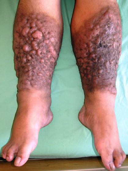
- Multiple waxy, indurated, nonpitting plaques and nodules with peau d’orange appearance and hyperpigmentation developing on bilateral legs.
Pretibial myxoedema is a rare manifestation of Graves’ disease that occurs in 0.5 to 4.3 per cent of patients. The classification of pretibial myxoedema includes the following four forms: non-pitting oedema; plaque; nodular; or elephantiasis. Only less than 1 per cent of patients with pretibial myxoedema develop elephantiasic form1. Characteristics of elephantiasic pretibial myxoedema include multiple nodular formation, massive oedema and skin hyperpigmentation. Differential diagnoses include secondary oedema resulting from chronic lymphatic obstruction or venous insufficiency, diabetic dermopathy, lichen amyloidosis, hypertrophic lichen planus, and rarely, pretibial epidermolysis bullosa. Elephantiasic pretibial myxedema is typically progressive and refractory to treatment2. Management for elephantiasic pretibial myxoedema remains a therapeutic challenge.
References
- Dermopathy of Graves’ disease (pretibial myxedema). Review of 150 cases. Medicine (Baltimore). 1994;73:1-7.
- [Google Scholar]
- Dermopathy of Graves’ disease (pretibial myxedema): long-term outcome. J Clin Endocrinol Metab. 2002;87:438-46.
- [Google Scholar]


