Translate this page into:
Development of immunodetection system for botulinum neurotoxin serotype E
For correspondence: Dr Ponmariappan Sarkaraisamy, Biotechnology Division, Defence Research Development & Establishment, Jhansi Road, Gwalior 474 002, Madhya Pradesh, India e-mail: pons@drde.drdo.in
-
Received: ,
This is an open access journal, and articles are distributed under the terms of the Creative Commons Attribution-NonCommercial-ShareAlike 4.0 License, which allows others to remix, tweak, and build upon the work non-commercially, as long as appropriate credit is given and the new creations are licensed under the identical terms.
This article was originally published by Medknow Publications & Media Pvt Ltd and was migrated to Scientific Scholar after the change of Publisher.
Abstract
Background & objectives:
Botulism, a potentially fatal paralytic illness, is caused by the botulinum neurotoxins (BoNTs) secreted by Clostridium botulinum. It is an obligate anaerobic, Gram-positive, spore-forming bacterium. BoNTs are classified into seven serotypes based on the serological properties. Among these seven serotypes, A, B, E and, rarely, F are responsible for human botulism. The present study was undertaken to develop an enzyme-linked immunosorbent assay (ELISA)-based detection system for the detection of BoNT/E.
Methods:
The synthetic gene coding the light chain of BoNT serotype E (BoNT/E LC) was constructed using the polymerase chain reaction primer overlapping method, cloned into pQE30UA vector and then transformed into Escherichia coli M15 host cells. Recombinant protein expression was optimized using different concentrations of isopropyl-β-D-1-thiogalactopyranoside (IPTG), different temperature and the rBoNT/E LC protein was purified in native conditions using affinity column chromatography. The purified recombinant protein was checked by sodium dodecyl sulphate-polyacrylamide gel electrophoresis (SDS-PAGE) and further confirmed by western blot and matrix-assisted laser desorption ionization-tandem time-of-flight (MALDI-TOF). Polyclonal antibodies were generated against rBoNT/E LC using Freund's adjuvant in BALB/c mice and rabbit. Sandwich ELISA was optimized for the detection of rBoNT/E LC and native crude BoNT/E, and food matrix interference was tested. The developed antibodies were further evaluated for their specificity/cross-reactivity with BoNT serotypes and other bacterial toxins.
Results:
BoNT/E LC was successfully cloned, and the maximum expression was achieved in 16 h of post-induction using 0.5 mM IPTG concentration at 25°C. Polyclonal antibodies were generated in BALB/c mice and rabbit and the antibody titre was raised up to 128,000 after the 2nd booster dose. The developed polyclonal antibodies were highly specific and sensitive with a detection limit about 50 ng/ml for rBoNT/E LC and 2.5×10³ MLD50 of native crude BoNT/E at a dilution of 1:3000 of mouse (capturing) and rabbit (revealing) antibodies. Further, different liquid, semisolid and solid food matrices were tested, and rBoNT/E LC was detected in almost all food samples, but different levels of interference were detected in different food matrices.
Interpretation & conclusions:
There is no immune detection system available commercially in India to detect botulism. The developed system might be useful for the detection of botulinum toxin in food and clinical samples. Further work is in progress.
Keywords
Biowarfare agents
botulinum neurotoxins
botulism
polyclonal antibody
recombinant protein
Botulism is a neuroparalytic disease caused by the neurotoxins secreted by Clostridium botulinum, which is an obligate anaerobic spore-forming bacterium. Botulinum neurotoxins (BoNTs) are classified into seven serotypes designated from A to G based on the serological properties1. BoNTs are initially synthesized as a single inactive polypeptide chain of 150 kDa which upon proteolytic cleavage is converted into active di-chain consisting of heavy chain (HC) of 100 kDa and light chain (LC) of 50 kDa joined together via disulphide linkage. C-terminal domain of HC is responsible for binding of neurotoxin to the targeted neuronal cell surface followed by receptor-mediated endocytosis. The toxins enter into the neuronal cell cytoplasm and LC cleaves its specific SNARE (soluble N-ethylmaleimide-sensitive factor attachment protein receptor) proteins involved in docking and fusion of acetylcholine neurotransmitter-containing synaptic vesicles. Cleavage of SNARE proteins blocks the release of acetylcholine in synapse region resulting in flaccid muscle paralysis. BoNT serotype A/C/E cleaves SNAP25 and BoNT serotype B/D/F/G cleaves vesicle-associated membrane protein (VAMP) and synaptobrevin2. BoNT types A, B, E and (rarely) F are mainly responsible for human botulism3.
Different botulism forms are reported depending on the mode of acquisition of toxin. Food-borne botulism (FBB) results from the ingestion of toxin contaminated food4. Wound botulism is caused by the organisms that multiply and produce toxin in a contaminated wound. Infant botulism (IB) occurs due to the endogenous production of toxin by C. botulinum after colonization in the intestine of infants5. IB cases were reported in the region of Qinghai-Tibet plateau of Northwest China caused by C. butyricum6. IB cases were also reported which were caused due to BoNT/E produced by C. butyricum isolated from tank water having pet terrapins7. FBB was first reported in India in 1998 from a residential school of rural Gujarat. Out of 310 students, 34 developed symptoms, 31 recovered after treatment and three students died mainly caused by BoNT/E-producing C. butyricum8.
BoNTs are the most toxic known substances with an estimated human lethal dose of 90-150 ng when injected intravenously or intramuscularly, 700-900 ng by inhalation and 70 μg orally9. Due to extreme toxicity of BoNTs, need for prolonged intensive care among affected persons and relative ease of production and transport, BoNTs can be used as bioterrorism agents10. At present, the mouse bioassay11 is the most reliable method for the detection of botulinum toxin in serum or food samples. Although the method is very sensitive and specific, but it is associated with a large number of drawbacks such as need for a large number of animals along with laboratory expertise and time consuming nature requiring at least four days for getting results. In addition, ethical issues are associated with the use of animals in laboratory. Hence, it is necessary to replace mouse bioassay with equally sensitive alternative assays1213. So far, in India, no detection system was available commercially against BoNT type E, so the present study was designed to develop an ELISA-based detection system against BoNT type E.
Material & Methods
This study was carried out in the Biotechnology division of Defence Research and Development Establishment (DRDE), Gwalior, India, from August 2015 to July 2016.
Synthetic botulinum neurotoxin serotype E light chain (BoNT/E LC) gene construction and cloning: The synthetic gene (1324 base pairs) coding BoNT/E LC was constructed using polymerase chain reaction (PCR) primer overlapping method14. Further, commercially procured BoNT/E LC specific primers (Sigma Aldrich, Bengaluru), forward primer 5’TAT GCA CAT ATG CCA AAA ATT AAT AGT TTT AAT 3’ and reverse primer 5’ TAG CAG CTC GAG TCA CCA TTA TTT ATT TCG ATAC 3’, were used for PCR amplification of LC gene. Amplification reaction was carried out for an initial denaturation at 95°C for 15 min followed by 35 cycles consisting of denaturation at 95°C for 30 sec, annealing at 56°C for one min 30 sec, extension at 72°C for two min and final extension at 72°C for 10 min in a T-gradient thermal cycler (Biometra, Germany). The amplified PCR product was checked in one per cent low melting agarose gel electrophoresis and further purified using DNA purification kit (Qiagen, Germany). The purified PCR product was quantified using Nanodrop1000 spectrophotometers (Thermo scientific, USA) and ligated into pre-linearized pQE 30 UA vector. Recombinant vector was transformed into chemically competent Escherichia coli M15 host cells. Transformants were then plated on Luria-Bertani (LB) agar plates containing kanamycin (30 μg/ml) and ampicillin (100 μg/ml). Plasmids were extracted from the chosen transformants using QIA miniprep kit (Qiagen, Germany) and screened for the presence of inserts using BoNT/E LC specific primers mentioned above and also checked for the orientation of insert using insert- and vector-specific primers (Sigma-Aldrich, Bengaluru).
Recombinant protein expression and localization: A single colony of positive transformant was inoculated into 5 ml LB broth containing kanamycin (30 μg/ml) and ampicillin (100 μg/ml) and incubated at 37°C overnight with constant shaking. Overnight grown culture was further inoculated into 50 ml super broth medium containing kanamycin (30 μg/ml) and ampicillin (100 μg/ml) and grown at 37°C with constant shaking till the optical density at 600 nm reaches 0.6-0.8 and the cells were then induced at different concentrations (0.25, 0.5 and 0.75 mM) of isopropyl-β-D-1-thiogalactopyranoside (IPTG) for the expression of the recombinant protein at two different temperatures (20 & 25°C) for 16-18 h. After induction, culture was centrifuged at 8000 ×g for 10 min at 4°C. Cell pellets were resuspended in phosphate-buffered saline (PBS, 50 mM NaH2PO4, 300 mM NaCl, pH 7.4) and lysozyme was added at 1 mg/ml concentration, mixed well and incubated on ice for 30 min followed by sonication on ice for 10 sec pulse on with 10 sec pause for 5-10 min. Cell lysate was centrifuged at 10,000 ×g at 4°C for 30 min. After that, the cell pellets as well as supernatant (lysate) were run in sodium dodecyl sulphate-polyacrylamide gel (SDS-PAGE) to check the localization of the expressed recombinant protein15. For un-induced control, 1 ml culture aliquot was taken out aseptically prior to IPTG induction.
Purification of recombinant protein: Ni-NTA (nickel-nitrilotriacetic acid) metal affinity chromatography was used for purification of rBoNT/E LC protein in native conditions. Clear cell lysate (4 ml) was mixed with 1 ml 50 per cent Ni-NTA slurry (Qiagen, Germany) on ice by shaking at a rotary shaker for one hour. Cell lysate-slurry mixture was loaded into the column, allowed the mixture to settle down and flow through was collected. Column was washed with wash buffers containing different concentrations of imidazole (50 mM NaH2PO4, 300 mM NaCl, 10/20/75/100 mM imidazole)16. Different wash fractions were collected and stored for SDS-PAGE analysis. The column was filled with the elution buffer (50 mM NaH2PO4, 300 mM NaCl, 250 mM imidazole). Elute fractions were collected and analyzed by SDS PAGE. All the elute fractions were pooled together, dialyzed against PBS (137 mM NaCl, 2.7 mM KCl, 8 mM Na2HPO4 and 2 mM KH2PO4) and concentrated using amicon filter (Millipore, USA). Protein concentration was determined using Bradford protein assay kit (Bio-rad, USA).
Western blot analysis: rBoNT/E LC protein is tagged with six histidine residues. Commercially available anti-His monoclonal antibody conjugated with Horseradish peroxidase (HRP) enzyme (Sigma, USA) was used to confirm the presence of purified tagged protein. Purified rBoNT/E LC protein was run in 12 per cent SDS-PAGE, along with prestained molecular weight marker (MBI Fermentas, Canada) and electrophoretically transferred to the polyvinylidene fluoride (PVDF) membrane (Millipore, USA). The membrane was incubated in 3 per cent skimmed milk (dissolved in 1X PBS) at 4°C for overnight. The PVDF membrane was washed three times with PBS containing 0.05 per cent Tween-20 (PBST) and one time with PBS and then incubated in 1:2000 dilution of mouse anti-His HRP -conjugated monoclonal antibody (Sigma-Aldrich, USA) for 1 h with gentle shaking on the rotary shaker. Then, the membrane was again washed three times with PBST and one time with PBS. Finally, protein bands were visualized by incubating the membrane in DAB (3,3’-diaminobenzidine) (Sigma-Aldrich, USA) substrate.
Matrix-assisted laser desorption ionization-tandem time-of-flight (MALDI-TOF-TOF) analysis: Protein band was excised from the gel using a fresh scalpel blade. Gel piece was washed three times with proteomic-grade distilled water, destained, reduced and alkylated and then digested with trypsin using Montage in-Gel digest kit (Millipore, USA) as per manufacturer's instructions. Protein was identified using a matrix-assisted laser desorption ionization-tandem time-of-flight (MALDI-TOF-TOF) mass spectrometer.
Antibody generation: Animals for experiment were maintained in Biotechnology division, DRDE (Gwalior, India). The animal experiments were approved by the Institutional Ethics Committee, DRDE. BALB/c mice (15-25 g) and Australian white rabbits (1.3-2.0 kg) were selected for immunization. A group of five BALB/c mice were immunized intramuscularly with 10 μg of rBoNT/E LC protein emulsified with Freund's complete adjuvant (Sigma-Aldrich, USA) on day 0, followed by 20 and 30 μg of purified recombinant protein emulsified with Freund's incomplete adjuvant (Sigma-Aldrich, USA) as booster doses on days 14 and 28. Australian white rabbits were immunized intramuscularly with 100 μg of purified recombinant protein emulsified with Freund's complete adjuvant on day 0 and with 200 and 400 μg of purified recombinant protein emulsified in Freund's incomplete adjuvant as booster doses on days 14 and 28. Mice were bled with glass capillary through retro-orbital puncture and blood was taken from the heart of the rabbit prior to primed dose and after the 2nd booster dose. Blood was incubated at 37°C for 30 min and then at 4°C for 1 h, and pre-immune and hyper-immune serum samples were collected by centrifuging the blood samples at 5000 ×g for 10 min to remove blood debris. All serum samples were stored at −80°C for further use.
Antibody titre determinations by ELISA: Serum was used for the antibody titre determination raised against rBoNT/E LC protein. Microtitre ELISA plates (Nunc-Immuno plate with Maxisorp surface, Nunc, Denmark) were coated with 500 ng/well of rBoNT/E LC protein in coating buffer (0.05 M carbonate-bicarbonate buffer pH 9.6) and incubated at 4°C overnight. Plates were blocked with 200 μl of 3 per cent bovine serum albumin (BSA) (Sigma, USA) made in PBS for 2 h at 37°C. Plates were washed three times with PBST and twice with PBS. Mouse anti-rBoNT/E LC and rabbit anti-rBoNT/E LC test serum samples were two-fold serially diluted in PBS containing 1 per cent BSA starting from 1:1000 to 1:512000 and incubated in triplicate (100 μl/well) at 37°C for one hour. Plates were washed three times with PBST and twice with PBS. Rabbit anti-mouse IgG-HRP (Dako, USA) and goat anti-rabbit IgG-HRP (Dako) were diluted in PBS (1:2000) containing one per cent BSA and added to the wells (100 μl/well) and incubated at 37°C for one hour. The plates were washed as above and then the plates were developed at 37°C for 15 to 20 min by adding H2O2-containing 2, 2’-azino-bis (3-ethylbenzo-thiazoline-6-sulphonic acid) diammonium salt (ABTS) substrate. Absorbance was measured at 410 nm using an ELISA plate reader (Biotek, USA). The means of absorbance values and standard deviation (SD) for each triplicate samples were calculated. Hyper-immune serum sample dilutions at which the absorbance value was two times the cut-off value were considered to be the ELISA endpoint. The cut-off value was determined as the mean specific optical density plus three times the SD for pre-immune serum assayed at a dilution of 1:1000.
Sandwich ELISA: Checkerboard titration was done after the final booster dose of antigen to find the optimum dilutions of rabbit and mouse anti-rBoNT/E LC serum, and 1:3000 dilutions were found suitable for sandwich ELISA15. The raised mouse anti-rBoNT/E LC antibody was diluted to 1:3000 in coating buffer and used to coat the polystyrene wells (100 μl/well) in triplicate and incubated overnight at 4°C. Next morning, the plate was washed three times with PBST and two times with PBS, and the remaining sites of adsorption were blocked by adding 3 per cent BSA (200 μl/well) and the plate was further incubated for 1 h at 37°C. The plate was washed three times with PBST and two times with PBS. Purified rBoNT/E serially diluted (100 μg/ml to 0.0001 μg/ml) in PBS containing one per cent BSA was added (100 μl/well), similarly native crude BoNT/E was used from 5×10³ to 0.6×10³ MLD50(50% lethal dose) of mice (100 μl/well) and incubated for 1 h at 37°C. After that, the plate was washed three times with PBST and two times with PBS. The plate was incubated with raised rabbit anti-rBoNT/E LC antibody (100 μl/well) at the dilution of 1:3000 and incubated for 1 h at 37°C. The plate was washed as mentioned above and incubated at 37°C for 1 h with the HRP-conjugated goat anti-rabbit IgG at a dilution of 1:2000 (Dako, USA). The wells were washed three times with PBST and two times with PBS. The plate was developed by adding H2O2-containing ABTS substrate at 37°C for 15 to 20 min. Absorbance was measured at 410 nm using an ELISA plate reader. The cut-off value for assay was calculated as the mean specific optical density plus three times the SD of the well having no antigen. Concentration of the rBoNT/E LC protein at which the absorbance value was two times the cut-off value (A410 using the well having no antigen) was considered to be the ELISA endpoint (detection limit).
Evaluation of food matrix interference in the detection of rBoNT/E LC: In liquid food matrices (orange juice, whole milk, coke samples), rBoNT/E LC protein was artificially spiked at a concentration of 1 μg/ml, mixed by vortexing and incubated at 25°C for 30 min. Samples were centrifuged at 7000 ×g for 30 min to remove any solid particle. Supernatant was diluted 1:1 with PBS in an Eppendorf tube to get the final concentration of 0.5 μg/ml, mixed thoroughly and used for sandwich ELISA. In case of solid food matrices (chicken, meat, dal, honey), 2 g food sample was spiked with rBoNT/E LC-purified protein, and incubated at 25°C for 30 min. Following incubation, PBS was added to get the final concentration of 0.5 μg/ml and further homogenized using bench-top Stomacher®(Seward, UK). After homogenization, samples were centrifuged at 7000 ×g for 30 min to remove any solid particle. The supernatant was used for sandwich ELISA to find any hindrance caused by any matrices in the detection of rBoNT/E LC protein17, and the recovery of rBoNT/E from spiked food samples is described as a percentage recovery by comparing to the standard value of rBoNT/E spiked in PBS (1% BSA).
The specificity and cross-reactivity of the raised polyclonal antibody were checked using 100 μg/ml of TCA (trichloroacetic acid)-precipitated crude culture supernatants of C. botulinum serotypes [type A (DB121CLB08), B (DB123CLB11), E (DB113CLB01) and F (DB116CLB04)], closely related other clostridial species such as C. tetani (CRI, Kasauli) and C. sordellii ATCC 9714, C. perfringens ATCC-13124, cell lysates of Staphylococcus aureus and Shigella dysenteriae, 10 μg/ml of purified recombinant antigens such as rBoNT/A LC, rBoNT/B LC, PA (protective antigen) and LF (lethal factor) anthrax toxin, epsilon toxin (Etx) of C. perfringens, ricin and abrin toxin, which were obtained from Biotechnology division, DRDE, Gwalior.
Results
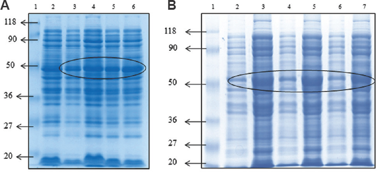
Synthetic gene construction, cloning and expression of rBoNT/E LC: Primers were designed to construct BoNT/E LC and synthesized commercially. Using the synthesized primers, the synthetic gene of 1324 bp was constructed by PCR amplification (Fig. 1). The constructed synthetic gene fragment was confirmed by sequencing as well as multiple sequence alignment with the nucleotide sequence of LC of standard C. botulinum type E available in NCBI database (https://www.ncbi.nlm.nih.gov/nucleotide/AB082519.1). The multiple sequence alignment results revealed that the constructed sequence showed 100 per cent similarity with standard BoNT/E LC. The synthetic gene sequence was submitted as GenBank with Accession No. HM008255. After confirmation, PCR product was gel purified and cloned into pQE30UA vector and then transformed into E. coli M15 cells. Chimeric transformants were selected on kanamycin and ampicillin containing LB agar plates. Some transformants were selected and plasmid was isolated to check the presence of insert using BoNT/E LC-specific forward and reverse primers. Recombinant protein expression conditions were optimized using 0.25, 0.5, 0.75 mM IPTG concentration as well as at two different temperatures (20 & 25°C), and the maximum yield was obtained at absorbance (OD600) 0.6-0.8 with 0.5 mM IPTG induction for 16-18 h at 25°C and the results are shown in (Fig. 2A and B).

- PCR amplification of botulinum neurotoxin serotype E light chain gene. Lane 1, molecular weight marker (bp); lanes 2-4, Botulinum neurotoxin serotype E light chain gene.

- SDS-PAGE for protein expression of Botulinum neurotoxin serotype E light chain clone (A) induced at 20°C; lane 1, protein molecular weight marker (kDa); lane 2, Escherichia coli M15 host cells; lane 3, uninduced clone; lanes 4-6, induced with 0.25, 0.5, 0.75 mM isopropyl-β-D-1-thiogalactopyranoside, (B) induced at 25°C; lane 1, protein molecular weight marker (kDa); lanes 2,4,6, uninduced clone; lane 3,5,7, induced with 0.25, 0.5, 0.75 mM isopropyl-β-D-1-thiogalactopyranoside. Circle indicates 50 kDa protein. SDS-PAGE, sodium dodecyl sulphate-polyacrylamide gel electrophoresis.
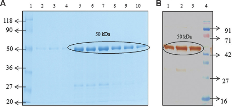
Purification and confirmation of rBoNT/E LC protein: After induction, cells were harvested at 8000 ×g for 10 min at 4°C. Localization of the expressed protein was done by lysing the cell pellets in PBS containing 1 mg/ml lysozyme, and the study results revealed that the recombinant protein was found in soluble form. Therefore, efforts were made to purify protein in native conditions using Ni-NTA affinity chromatography with various combinations of wash buffers, and finally rBoNT/E LC protein was purified up to about 98 per cent homogeneity and the results are illustrated in (Fig. 3A). Further protein was confirmed by Western blotting using anti-His antibodies (Fig. 3B) as well as by MALDI-TOF-TOF. The purified recombinant protein showed similarity with catalytic domain of C. botulinum type E standard culture.

- (A) SDS-PAGE. Lane 1, protein molecular weight marker (kDa); lanes 2 to 10, purified protein. (B) Western blot. Lanes 1 to 3, purified protein; lane 4, protein molecular weight marker (kDa). Circle indicates Botulinum neurotoxin (50 kDa) protein.
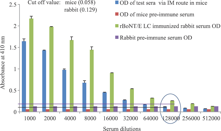
Polyclonal antibody generation: The hyper-immune serum was raised in mice and rabbits using rBoNT/E LC. Groups of mice and rabbit were injected with the antigen intramuscularly. Serum samples were collected on the seventh day after the 2nd booster dose and compared using ELISA, with pre-immune serum used as a negative control. Indirect ELISA was done to check the antibody titre by coating the wells with the 500 ng purified protein per well. Antibody titre was raised up to a serum dilution of 128,000 in both mice and rabbits after the 2nd booster dose of antigen with the cut-off value of 0.058 and 0.129, respectively, as shown in Fig. 4.

- Antibody titre raised against rBoNT/E light chain-soluble protein in mice (intramuscular route) and rabbit (intramuscular route). Optical density values represented are mean±standard deviation of triplicate determinations. Circle indicates antibody titre raised up to a serum dilution of 1:128000 in both mice and rabbit.
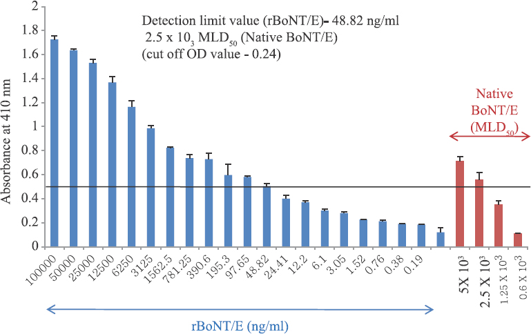
Evaluation of detection limit: Detection sensitivity of the sandwich ELISA was tested using rBoNT/E LC protein in the concentration range from 100 to 0.0001 μg/ml and native crude BoNT/E from 5×10³ to 0.6×10³ MLD50 as shown in Fig. 5. Antibody raised against rBoNT/E LC could detect about 50 ng/ml rBoNT/E LC and 2.5×10³ MLD50 of native crude BoNT/E with the cut-off value of 0.24. No cross-reactivity was found with culture supernatants of C. botulinum types A, B and F, C. tetani, C. sordellii, C. perfringens, S. aureus, S. dysenteriae as well as recombinant antigens such as rBoNT/A LC, rBoNT/B LC, Etx, PA-LF, ricin and abrin (results not shown).

- Minimum detection limit of botulinum neurotoxin type serotype E by sandwich ELISA. OD values represented are mean±standard deviation of triplicate determinations. Horizontal line indicates the detection limit (absorbance value two times of the cut-off value).
Discussion
Botulism, a neuroparalytic illness, is most commonly caused by ingestion of food containing preformed neurotoxin types A, B, E and F. Botulism is often fatal if untreated. Paralysis in case of botulism can be long-lasting, with concomitant requirement of supportive care. The onset of paralytic manifestations occurs from a few hours to over a week after intoxication, but in majority of cases, paralysis occurs in 12 to 72 h18. Definitive diagnosis requires identification of toxin in serum, gastric aspirate and stool. Mouse bioassay has been considered as the most reliable method for detection of BoNTs in serum or food samples, but it is time-consuming as it takes up to four days to get final results. The clinical diagnosis of botulism depends on clinical symptom development, thus limiting the effectiveness of antitoxin as it cannot reverse the existent paralysis (since antitoxin cannot cross the nerve membrane to neutralize the toxin inside the poisoned cells). A sensitive and rapid diagnosis of BoNTs is critical, if preventive measures are to be effectively employed. Handling large quantities of BoNTs poses serious health risks. To avoid such problems and to attain high yield of protein, synthetic gene approach was applied. Hence, in the present study, primers were designed and synthesized commercially for the construction of BoNT/E LC. Using these primers, LC was constructed by PCR and was confirmed by sequencing as well as multiple sequence alignment with the nucleotide sequence of LC of standard C. botulinum type E available in NCBI database, cloned into pQE 30UA vector and then transformed into E. coli M15 cells. Expression conditions were optimized and the maximum yield was obtained in overnight induction with 0.5 mM IPTG at 25°C. Recombinant protein was purified in native conditions using imidazole buffer and the maximum yield of the recombinant protein was about 10-12 mg/l. Agarwal et al19 cloned the BoNT/E LC in pET −9C vector and expressed in E. coli BL21DE3. The recombinant proteins purified in native forms and when stored at 4-10°C, the proteins undergo auto-proteolysis. The present study results revealed that, up to two week storage, there was no precipitation of the recombinant BoNT/ E LC protein at 4°C. The thermal stabilization of BoNT/E LC recombinant protein was achieved by phosphorylation of tyrosine amino acid at position 67 using site-directed mutagenesis which resulted in increased stability without compromising its biological activity20. Further structural analysis of BoNT/E LC proteins reveals that mutation of amino acid at position 212 (Glu to Gln) leads to loss of the catalytic activity of the proteins. The study also suggested that genetic modification of amino acid at position 212 (Glu to Ala) provides a novel vaccine candidate molecule for botulism21.
Further to raise antibodies, mice and rabbits were immunized with rBoNT/E LC protein mixed with Freund's adjuvant22. In the present study, a high antibody titre of 128,000 was obtained after the 2nd booster dose of antigen. The BoNT serotypes exhibit 30-60 per cent identity; however, serotype-specific antisera have been reported to elicit little or no cross-reactivity23. Similarly, our raised antibody did not show any cross-reactivity with other botulinum serotypes, other clostridial species and bacterial toxins. Antibody raised against rBoNT/E LC could detect about 50 ng/ml of rBoNT/E and 2.5×10³ MLD50 of native crude BoNT/E. Numerous ELISA formats with several modifications have been developed to enhance assay sensitivity, but these modifications require more specialized equipment. A sensitive ELISA was developed for the detection of BoNTs A, B and E using signal amplification via enzyme-linked coagulation assay with a detection limit of 5-10 pg/ml equivalents to mouse bioassay24. This method needs expensive reagents, requires many tedious steps for washing and complicated amplification process, sample throughput is low, testing time is relatively long and is difficult for automation. Sharma et al17 used digoxigenin-labelled antibodies which can detect up to 60 pg/ml BoNT/A, 176 pg/ml BoNT/B, 163 pg/ml BoNT/E and 117 pg/ml BoNT/F in casein buffer.
Sensitivity is a key part of any diagnostic system. The desired sensitivity can be obtained if the sample does not have any interfering substances with the detection system. Serum and environmental samples hardly require any processing and can be used directly for detection, but the food samples require processing for the extraction of analyte to be used in detection. Here, we tested different liquid, semisolid and solid food samples, and rBoNT/E LC was detected in almost all food samples, but considerable variations were observed in the absorbance value at the same concentration of protein. Differences in the absorbance values can be due to the matrix interference as each food sample has a different biochemical composition.
In conclusion, a simple and sensitive ELISA technique was developed that might be useful to test food samples in preliminary screening even though more validation is still needed with BoNT/E using a large number of samples before it can be utilized as a screening system in field conditions.
Financial support & sponsorship: The first author (RS) acknowledges University Grant Commission (UGC), India for financial support as Senior Research Fellow (SRF).
Conflicts of Interest: None.
References
- Botulinum neurotoxins: Biology, pharmacology, and toxicology. Pharmacol Rev. 2017;69:200-35.
- [Google Scholar]
- Subunit vaccine against the seven serotypes of botulism. Infect Immun. 2008;76:1314-8.
- [Google Scholar]
- Report of two unlinked cases of infant botulism in the UK in October 2007. J Med Microbiol. 2009;58:1601-6.
- [Google Scholar]
- Infant botulism due to C. butyricum type E toxin: A novel environmental association with pet terrapins. Epidemiol Infect. 2015;143:461-9.
- [Google Scholar]
- Outbreak of suspected Clostridium butyricum botulism in India. Emerg Infect Dis. 1998;4:506-7.
- [Google Scholar]
- Botulinum toxin as a biological weapon: Medical and public health management. JAMA. 2001;285:1059-70.
- [Google Scholar]
- Development of immunodetection system for botulinum neurotoxin type B using synthetic gene based recombinant protein. Indian J Med Res. 2011;134:33-9.
- [Google Scholar]
- Mouse bioassay versus Western blot assay for botulinum toxin antibodies: Correlation with clinical response. Neurology. 1998;50:1624-9.
- [Google Scholar]
- Development of novel assays for botulinum type A and B neurotoxins based on their endopeptidase activities. J Clin Microbiol. 1996;34:1934-8.
- [Google Scholar]
- Electrochemical immunosensor for botulinum neurotoxin type-E using covalently ordered graphene nanosheets modified electrodes and gold nanoparticles-enzyme conjugate. Biosens Bioelectron. 2015;69:249-56.
- [Google Scholar]
- Effect of adjuvants on antibody titer of synthetic recombinant light chain of botulinum neurotoxin type B and its diagnostic potential for botulism. J Microbiol Biotechnol. 2011;21:719-27.
- [Google Scholar]
- Development of ELISA based detection system for lethal toxin of Clostridium sordellii. Indian J Med Res. 2013;137:1180-7.
- [Google Scholar]
- Expression, purification and development of neutralizing antibodies from synthetic BoNT/B LC and its application in detection of botulinum toxin serotype B. Protein Pept Lett. 2012;19:288-98.
- [Google Scholar]
- Detection of type A, B, E, and F Clostridium botulinum neurotoxins in foods by using an amplified enzyme-linked immunosorbent assay with digoxigenin-labeled antibodies. Appl Environ Microbiol. 2006;72:1231-8.
- [Google Scholar]
- Microbiological, biological, and chemical weapons of warfare and terrorism. Am J Med Sci. 2002;323:326-40.
- [Google Scholar]
- Cloning, high level expression, purification, and crystallization of the full length Clostridium botulinum neurotoxin type E light chain. Protein Expr Purif. 2004;34:95-102.
- [Google Scholar]
- Thermal stabilization of the catalytic domain of botulinum neurotoxin E by phosphorylation of a single tyrosine residue. Biochemistry. 2001;40:2234-42.
- [Google Scholar]
- Structural analysis of botulinum neurotoxin type E catalytic domain and its mutant Glu212->Gln reveals the pivotal role of the Glu212 carboxylate in the catalytic pathway. Biochemistry. 2004;43:6637-44.
- [Google Scholar]
- Comparative study of immunological and structural properties of two recombinant vaccine candidates against botulinum neurotoxin type E. Iran Biomed J. 2012;16:185-92.
- [Google Scholar]
- Enzyme-linked immunosorbent assay and enzyme-linked coagulation assay for detection of Clostridium botulinum neurotoxins A, B, and E and solution-phase complexes with dual-label antibodies. J Clin Microbiol. 1994;32:105-11.
- [Google Scholar]


