Translate this page into:
Construction & establishment of two minigenome rescue systems for Chandipura virus driven by recombinant vaccinia virus expressing T7 polymerase
Reprint requests: Dr. Prasenjit Chakraborty, Department of Biotechnology, University of Calcutta, 35, Ballygunge Circular Road, Kolkata 700 019, West Bengal, India e-mail: cprasen8@gmail.com
-
Received: ,
This is an open access article distributed under the terms of the Creative Commons Attribution-NonCommercial-ShareAlike 3.0 License, which allows others to remix, tweak, and build upon the work non-commercially, as long as the author is credited and the new creations are licensed under the identical terms.
This article was originally published by Medknow Publications & Media Pvt Ltd and was migrated to Scientific Scholar after the change of Publisher.
Abstract
Background & objectives:
Chandipura virus (CHPV) is an emerging pathogenic rhabdovirus with a high case fatality rate. There are no reports of a minigenome system for CHPV, which could help its study without having to use the infectious agent. This study was, therefore, undertaken for the establishment of T7 polymerase-driven minigenome system for CHPV.
Methods:
The minigenome rescue system for CHPV consists of three helper plasmids expressing the nucleocapsid protein (N), phosphoprotein (P) and large protein (L) based on a recombinant vaccinia virus expressing bacteriophage T7 polymerase (vTF7-3). The minigenome construct is composed of a reporter gene, flanked by the non-coding regions of CHPV. Two minigenomes were constructed in an antigenome or complimentary sense, expressing luciferase or green fluorescent protein (GFP). The minigenome system was evaluated by co-transfection of the minigenome construct and three helper plasmids into CV-1 cells and analysis of the reporter gene activity.
Results:
All the helper proteins were expressed from the helper plasmids confirmed by Western blotting. Expression of reporter genes was observed from both the GFP and luciferase-based minigenomes. Green fluorescence could be visualized directly in live CV-1 cells. Luciferase activity was found to be significantly different from control.
Interpretation & conclusions:
The results showed that the helper plasmids provided all the necessary viral structural proteins required for the production of minigenome mRNA template, which in turn could rescue the expression of reporter genes. Thus, these minigenomes can be applied to mimic the manifestation of CHPV life cycle.
Keywords
Chandipura virus
CV-1 cell line
green fluorescent protein
luciferase
minigenome
recombinant vaccinia virus
T7 polymerase
The large group of negative-sense RNA viruses includes highly prevalent human pathogens such as rabies virus, respiratory syncytial virus, Ebola virus, Marburg virus, influenza virus and Chandipura virus (CHPV). CHPV is an emerging tropical pathogen with a case fatality rate of 55-75 per cent123456. Sequence analysis of CHPV has confirmed a genome organization similar to that of vesicular stomatitis virus (VSV) i.e., a non-segmented negative strand RNA genome encoding five transcription units separated by intragenic spacer regions, ordered along the genome as N–P–M–G–L in the 3' to 5' sense. At the 3' end, CHPV genome RNA comprises a 49 nucleotides leader gene that is transcribed but remains untranslated. A short non-transcribed 79 nucleotides trailer sequence is present at the 5' end. The complete genome sequence comprising 11,119 nucleotides has been determined7. After entry into the cell, the viral polymerase starts transcribing the different mRNAs, and after accumulation of significant amount of different viral polypeptides, the polymerase uses the negative sense genome to synthesize the full-length positive sense RNA, which is then utilized for amplification of genome RNA by the same polymerase8. Due to these features and its close resemblance to VSV, CHPV has been grouped into the genus Vesiculovirus, under the virus order Mononegavirales and family Rhabdoviridae.
Inside a living cell, a minigenome can resemble the life cycle of a virus. In these systems, the viral coding genes are replaced with a reporter gene such as chloramphenicol acetyltransferase (CAT), green fluorescent protein (GFP) or luciferase. However, the non-coding regions are retained allowing them to serve as templates for the viral polymerase. In the case of CHPV, these signals are located within the genome termini called the leader and trailer elements910. The required viral proteins are provided through helper plasmids. Currently, no minigenome system is available using only the minimal leader and trailer sequences of CHPV and three helper plasmids. This study was conducted to establish a T7 polymerase-driven minigenome system for CHPV, which would be helpful for studying its replication and transcription quantitatively and thus, for screening and developing new antiviral drugs. Similar minigenomes have been constructed and reported in related viruses such as Crimean-Congo haemorrhagic fever virus11, Ebola virus12, Newcastle disease virus13 and rabies virus14.
Material & Methods
This study was conducted in the department of Biotechnology, University College of Science and Technology, University of Calcutta and department of Structural Biology and Bioinformatics, Indian Institute of Chemical Biology, Kolkata, India, during June 2011-October 2015.
Cell culture and viral infection
Monkey kidney cell line CV-1 cells (National Centre for Cell Science Repository, Pune) were cultured in minimum essential medium (MEM) supplemented with 10 per cent foetal bovine serum (FBS) (Gibco, Thermo Fisher Scientific, USA). The cells were frozen in 20 per cent FBS-containing MEM supplemented with five per cent cryoprotective agent dimethyl sulphoxide. Frozen cells were revived in 20 per cent FBS-containing MEM. Recombinant vaccinia virus expressing bacteriophage T7 polymerase (vTF7-3) was procured from American Type Culture Collection (ATCC), USA. All vTF7-3 infections were performed in MEM containing 2.5 per cent FBS (MEM-2.5). Just before use, an equal volume of vTF7-3 stock and 0.25 mg/ml trypsin were mixed and vortexed vigorously to break up any clumps of cells. If the clumps were still visible, the vortexed stock was chilled to 0°C and sonicated with a 30 sec pulse on ice.
Titration of vTF7-3 stocks by plaque assay
Expression of reporter gene in the CHPV minigenome system requires the presence of T7 polymerase in CV-1 cells. For this purpose, recombinant vaccinia virus expressing bacteriophage T7 polymerase (vTF7-3) was used. Prepared stocks of vTF7-3 were quantitated by plaque assay on monolayers of CV-1 cells. Serial dilutions of the trypsinized vTF7-3 stock were used to infect the CV-1 cell line. After two days of growth, the medium was removed and the cells were stained with crystal violet. Plaques appear as 1 to 2 mm diameter areas of diminished staining due to the retraction, rounding and detachment of infected cells. The viral titre used for subsequent experiments was calculated to be 3.2 × 10⁷ plaque-forming unit (pfu).
Helper plasmids construction
N and P gene cloned under T7 promoter in pET3a vector was kindly provided by Prof. Dhrubajyoti Chattopadhyay (Guha Centre for Genetic Engineering and Biotechnology, University College of Science, University of Calcutta, Kolkata). The cloning of N and P gene has been reported previously1516. The 6.3 kb L gene of CHPV was subcloned into the expression vector pRSFDuet-1. The L gene originally cloned in pGEM-T Easy vector (kindly provided by Prof. Dhrubajyoti Chattopadhyay) was amplified by polymerase chain reaction (PCR)17 with primers (Integrated DNA Technologies, Inc. USA), specific for the gene: upstream (5'-AGACACAGTCGACATGGATCTCAACCCGG TGGATG-3') and downstream (5'-CAGACGCAGCGGCCGCTTAATCTACCATGCTCAG-3') having SalI and NotI restriction sites (underlined), respectively. The PCR products as well as the vector pRSFDuet-1 were digested by SalI and NotI and ligated with T4 DNA ligase to obtain the expression vector pRSFDuet-1-LC. All clones were verified by DNA sequencing1618.
Minigenome plasmids construction
To construct a T7 polymerase-driven luciferase or GFP-expressing CHPV minigenomes, a synthetic gene construct containing the following elements sequentially from the 5' end: hammerhead ribozyme (HAMRz), viral leader sequence, reporter gene sequence coding for either luciferase or GFP, viral trailer sequence, hepatitis delta virus antigenomic ribozyme (HDVRz) and T7 terminator was custom synthesized from GenScript (USA) and cloned into the T7 promoter-containing plasmid vector pBluescript II SK(+) at HindIII and XhoI restriction sites (Fig. 1). The minigenome with luciferase as the reporter gene was named as MGCL and the minigenome construct with GFP was named as MGCG. The clones were verified by DNA sequencing.
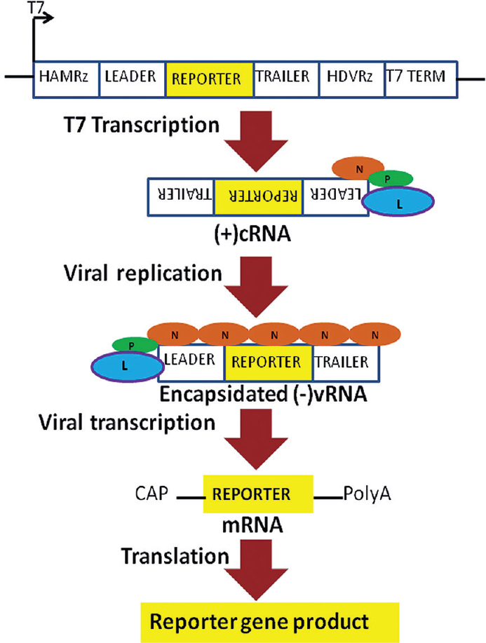
- Schematic representation of Chandipura virus minigenome constructs and reporter gene expression; The leader and trailer regions of Chandipura virus along with the reporter gene sequence is flanked by the hammerhead ribozyme (HAMRz) and the hepatitis delta virus ribozyme (HDVRz). The T7 RNA polymerase promoter (T7 on a bent arrow) coming from the plasmid and the terminator (T7 TERM) are also shown. Transcription by T7 polymerase produces a positive sense complementary RNA (cRNA). cRNA interacts with the helper viral proteins (N, P and L) to form encapsidated negative-sense viral RNA (vRNA). Transcription of the vRNA by the helper viral proteins produces a 5' end guanosine diphosphate capped (CAP) and 3' end polyadenylated (PolyA) messenger RNA (mRNA) leading to reporter gene expression.
Immunoblotting
Whole cell lysate from vTF7-3 infected and plasmid DNA transfected cells were resolved on a six per cent (for L protein) or 10 per cent (for N and P protein) polyacrylamide gel. When electrophoresis was complete, the gel sandwich was disassembled and the stacking gel was removed. The protein samples were transferred from gel to membrane with the help of the Trans-Blot Turbo apparatus (Bio-Rad, India), mini format containing 0.2 μM nitrocellulose membrane. The gel was stained for total protein with Coomassie blue [50% (v/v) methanol; 0.05% (w/v) Coomassie brilliant blue R-250; 10% (v/v) acetic acid and 40% H2O] to verify transfer efficiency. The membrane was blocked with 5 ml blocking buffer [5% non-fat dried milk diluted in Tris-buffered saline Tween-20 (TBST)] for one hour with agitation on an orbital shaker to reduce the background. The membrane was washed four times by agitating with 200 ml TBST, 10-15 min each time. The rabbit IgG primary antibody against respective viral proteins (Anti-N, Anti-P or Anti-L) was added at a dilution of typically 1:3000 in blocking buffer overnight with agitation on an orbital shaker. The membrane was washed again six times by agitating with 200 ml TBST, 10-15 min each time. The membrane was then treated with 1:5000 diluted goat anti-rabbit IgG (H+L) secondary antibody in TBST. After washing with TBST, the membrane was developed according to appropriate visualization protocol in ChemiDoc XRS+ system with Image Lab software (Bio-Rad, India).
Green fluorescent protein (GFP) minigenome assay
CV-1 cells grown in eight-well slide chamber to 95 per cent confluence were infected with vTF7-3 at a multiplicity of infection of 5 pfu/cell for 45 min at 37°C. Cell monolayers were washed with phosphate-buffered saline and transfected with 0.2 μg minigenome plasmid MGCG, 0.15 μg pET3a-NC plasmid encoding CHPV N protein, along with 0.1 μg of plasmid pET3a-PC expressing the CHPV P protein and 0.05 μg of plasmid pRSFDuet-1-LC expressing the CHPV L protein, using Lipofectamine LTX with Plus reagent (Thermo Fisher Scientific, India) following the manufacturer's instructions. At five hours post-transfection, the medium was changed with fresh MEM-2.5. Twenty four hours post-transfection, live cells were observed and the fluorescence was monitored under an Andor Spinning Disk live cell confocal microsope (Belfast, UK) using appropriate settings.
Luciferase minigenome assay
CV-1 cells grown in six-well tissue culture plates to 95 per cent confluence were infected with vTF7-3 at a multiplicity of infection of 5 pfu/cell for 45 min at 37°C. Cell monolayers were washed with phosphate-buffered saline and transfected with 2 μg MGCL, 1.5 μg pET3a-NC plasmid encoding CHPV N protein, along with 1 μg of plasmid pet3a-PC expressing the CHPV P protein and 0.5 μg of plasmid pRSFDuet-1-LC expressing the CHPV L protein, using Lipofectamine LTX with Plus reagent following the manufacturer's instructions. At five hours post-transfection, the medium was replaced with fresh MEM-2.5. Twenty four hours post-transfection, cells were harvested in Passive Lysis Buffer (Promega, India) and lysates were subjected to reporter gene assays using the luciferase assay system. Luciferase activity was measured with a Luminometer (PerkinElmer VICTOR X3, India) according to the manufacturer's instructions.
siRNA knockdown-rescue experiment
Expression of P protein from the helper plasmid pET3a-PC in the minigenome systems was knocked down using P-2 siRNA19. During transfection, 20 pmol of P-2 siRNA or its scrambled form PS-2 siRNA (Qiagen, India) was added to GFP minigenome. For luciferase minigenome, the amount of siRNA added was 100 pmol. The expression of minigenome systems was assayed as described above. For knockdown rescue, a P-2 siRNA-resistant P protein clone (Psiwt) was used as the helper plasmid instead of pET3a-PC within the minigenome systems. The construction of Psiwt has been reported19.
Statistical analysis
A two-tailed unpaired Student's t test was used to determine the significance of differences between treated and control groups. GraphPad PRISM 5.01 (GraphPad Software, USA) software was used for statistical analysis.
Results
Expression of helper plasmids
vTF7-3-mediated expression of the N, P and L proteins in CV-1 cells was confirmed by Western blotting with antibodies directed against the respective proteins (Fig. 2). Genes for all the three proteins were cloned under a T7 promoter and the recombinant plasmids (pET3a-NC, pET3a-PC and pRSFDuet-1-LC) were transfected into CV-1 cells following vTF7-3 infection. As a control, CV-1 cells were transfected with empty vectors (EVs) following vTF7-3 infection. The results indicated that the expression plasmids pet3a-NC, pet3a-PC and pRSFDuet-1-LC could be used for transient expression of exogenous viral N, P and L proteins ex vivo.
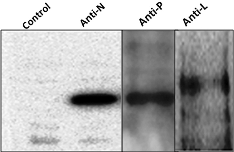
- Expression of N, P and L genes in CV-1 cells detected by immunoblotting; Whole-cell lysates from CV-1 cells infected with vTF7-3 and then transfected with pET3a-NC, pET3a-PC or pRSFDuet-1-LC plasmids were immunoblotted with antibodies specific for N, P or L proteins. As a control, cells infected with vTF7-3 and then transfected with empty vectors were used. Blots for each protein along with controls have been developed separately, but only one control is shown here.
Verification of the GFP minigenome system
To examine the efficiency of the minigenome rescue system for MGCG expressing the reporter gene GFP, CV-1 cells were infected with vTF7-3 at 5 pfu/cell. Forty five minutes post-infection, cells were co-transfected with MGCG, pET3a-NC, pET3a-PC and pRSFDuet-1-LC at a ratio of 4:3:2:1, respectively, with a total of 0.5 μg plasmid DNA. The applied ratio was based on the results obtained with minigenome system for the closely related VSV20. As a control, CV-1 cells infected with vTF7-3 were transfected with MGCG and EV, keeping the total amount of transfected plasmid DNA at 5 μg. A blank control was also done, where no plasmids were transfected. Live cells were then visualized under an Andor Spinning Disk live cell confocal microscope at 24 h post-transfection. The cells co-transfected with MGCG and helper plasmids showed GFP expression considerably more than those in the control groups at 24 h post-transfection (Fig. 3), demonstrating that a continuous production of T7-driven transcripts and their replication and transcription driven by viral proteins lead to an accumulation of reporter gene product (GFP) in the cells.
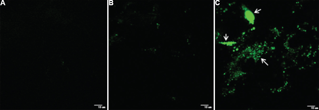
- Expression of green fluorescent protein from a positive-sense minireplicon of Chandipura virus; (A) CV-1 cells infected with vTF7-3 only. (B) CV-1 cells infected with vTF7-3 and transfected with MGCG and empty vector. (C) CV-1 cells infected with vTF7-3 and transfected with MGCG and helper plasmids expressing Chandipura virus N, P and L proteins (arrows).
Validation of the minigenome system expressing luciferase
To examine the expression of luciferase from MGCL, CV-1 cells were infected with vTF7-3 at 5 pfu/cell. Forty five minutes post-infection, the cells were transfected with MGCL, pET3a-NC, pET3a-PC and pRSFDuet-1-LC at a ratio of 4:3:2:1, respectively, with a total of 5 μg plasmid DNA (MGCL+NPL). As controls, CV-1 cells infected with vTF7-3 were transfected with MGCL and EV, MGCL and one or two helper plasmids (MGCL+N, MGCL+P, MGCL+L, MGCL+NP, MGCL+NL and MGCL+PL), keeping the total amount of transfected plasmid DNA at 5 μg. A blank control was also done, where no plasmids were transfected after infection with vTF7-3. The luciferase activity was significantly (P <0.005) higher in the co-transfected group containing MGCL and three helper plasmids expressing all the three CHPV proteins (MGCL+NPL) than in the control groups at 24 h post-transfection (Fig. 4).
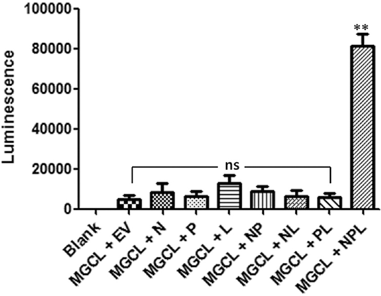
- Expression of luciferase from Chandipura virus minigenome; cells were treated with minigenome construct expressing luciferase (MGCL) and empty vector (EV) or one or two helper plasmids for controls (MGCL + N, MGCL + P, MGCL + L, MGCL + NP, MGCL + NL and MGCL + PL) as indicated in the graph and described in the text. Cells without any transfection of plasmids served as a blank control. MGCL + NPL indicate transfection with minigenome construct expressing luciferase and all three helper plasmids. The experiment was performed thrice and data points are shown as mean±SEM. **P <0.005 compared to blank control; ns, not significant.
Importance of CHPV P protein in minigenome expression
To prove that the activity of viral proteins expressed through helper plasmids is essential for proper expression of the established minigenome systems, a siRNA knockdown-rescue experiment was performed directed against P protein of CHPV with these systems. P-2 siRNA has already been shown to specifically downregulate CHPV P protein, which can be rescued by transient expression of P-2 siRNA-resistant P protein clone Psiwt19. Basically, both the GFP and luciferase minigenome systems were setup in the presence of P-2 siRNA (P-2+MGCG+NPL/ P-2+MGCL+NPL) or its scrambled form PS-2 (PS-2+MGCG+NPL/PS-2+MGCL+NPL). A blank control was also done with only vTF7-3 infection, against which the different minigenome systems were evaluated. Only P-2 siRNA was able to inhibit the expression of reporter genes in both GFP and luciferase minigenomes (Fig. 5). Minigenome systems knocked down with P-2 siRNA were rescued by Psiwt(P-2+MGCG+NPsiwtL/P-2+MGCL+NPsiwtL). Extraneous expression of a siRNA-resistant P protein clone could rescue the minigenome phenotype significantly as shown in both Fig. 5A and B (P <0.001).
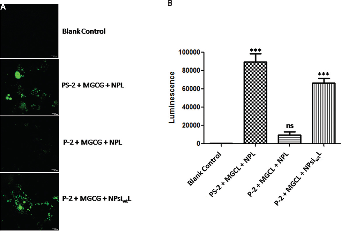
- Chandipura virus P protein is essential for the minigenomes; siRNA knockdown-rescue experiment was performed to show the importance of P protein for Chandipura virus minigenome expression. (A) Effect of siRNA knockdown and subsequent rescue on green fluorescent protein minigenome. CV-1 cells infected with vTF7-3 and transfected with the indicated siRNA and plasmids were visualized by confocal microscopy. (B) siRNA knockdown-rescue on luciferase minigenome. Luminescence was measured after differential treatment of luciferase minigenome as shown in the Figure. Result is expressed as mean±standard error of mean (n=3). ***P <0.001 compared to blank control; ns, not significant. P-2, siRNA against CHPV P protein; PS-2, scrambled form of P-2; MGCG, minigenome construct expressing GFP; MGCL, minigenome construct expressing luciferase; NPL, all three helper plasmid clones: pET3a-NC, pET3a-PC and pRSFDuet-1-LC; NPsiwtL, a P-2 siRNA-resistant P protein clone used as the helper plasmid, instead of pET3a-PC along with pET3a-NC and pRSFDuet-1-LC.
Discussion
Less than two decades ago, it was not possible to generate negative-sense RNA viruses21. Now, through a series of major scientific advances, minireplicon system is available for negative-sense RNA viruses and investigators have the tools to genetically engineer these viruses. The insights afforded by these advances can increase understanding of virus life cycles and clarify the mechanisms by which these viruses cause diseases. In 1994, Schnell et al22 reported for the first time generation of minigenome rescue system for rhabdoviruses. One year later, reverse genetic system was established for the prototype rhabdovirus VSV23. The importance of the PPPY motif in the amino-terminal part of VSV matrix protein and its interaction with dynamin for efficient virus budding was revealed using minigenome systems2425. Recombinant attenuated VSV strains have shown promising activity selectively against cancer cells26. Reverse genetics can also be applied to produce live-attenuated vaccines against the many diseases caused by negative-sense RNA viruses2728.
In the process of CHPV infection, the viral genome is transcribed by the RNP complex containing the N, P and L proteins. The RNP complex begins transcription at 3' end of the genome, with progressive attenuation at each gene boundary to result in decreasing amount of transcripts for genes that are distant from the entry site. The RNP complex was proposed to remain associated with the N-RNA template while transcribing the genomic template and reinitiate synthesis of downstream genes after termination. Transcription from downstream genes is dependent on termination of upstream genes and reinitiation, controlled by cis-acting start and stop signals8. The entire process of CHPV replication and transcription has been simulated in a minigenome system, where the N, P and L proteins are provided through helper plasmids. In the minigenome plasmid, hammerhead and HDV ribozyme sequences are placed adjacent to the T7 promoter and terminator, respectively, to generate precise positive-sense cRNA-like transcript, which follows the replication and transcription pattern of CHPV in CV-1 cells. T7 polymerase-driven expression of all the three proteins from the helper plasmids constituting the CHPV ribonucleoprotein complex in CV-1 cells was observed by immunoblotting.
GFP and firefly luciferase-mediated bioluminescence is used as a sensitive reporter assay for gene expression. Luciferase activity can also be used as a suitable quantitative tool to explore the response of the minigenome system under various conditions such as in response to antiviral agents. Assays with luciferase activity can give approximately 100-fold greater sensitivity than that achieved with the CAT assay29. In the minigenomes activity assays, it was found that the helper plasmids expressed the functional viral N, P and L proteins ex vivo to drive the replication and transcription of the minigenome, leading to expression of the reporter gene. The reporter gene activity of minigenome and all the three helper plasmids' co-transfected cells was significantly higher than the negative, blank controls and when one or two helper plasmids were co-transfected. Knockdown rescue on the minigenomes using siRNA directed against the P protein and a siRNA-resistant P protein clone has shown the specific requirement of the properly folded and active viral protein for appropriate minigenome expression. The low level of transcription observed in the negative control groups can be attributed to a vaccinia virus-encoded capping mechanism or some unknown eukaryotic promoter-like sequences in the minigenome.
In conclusion, two minigenome systems of CHPV were established which could be used to model its life cycle. Minigenomes were evaluated based on the detection of reporter gene molecules by different methods. Before carrying out further research, specific functions of the helper viral proteins within the minigenomes must be established. In future, another minigenome can be developed that resembles the genomic or negative-sense viral RNA, where replication is not a prerequisite for reporter gene expression. Apart from testing of antivirals against CHPV, the minigenome system can also be applied to answer many intriguing questions regarding the life cycle of this deadly pathogen. The molecular mechanism that allowed for a switch in the viral polymerase function from transcription to replication, popularly known as the transcription-replication switch, has remained obscure. Development of a replication-deficient minigenome could be of great help to unravel the mechanism of the switch.
Acknowledgment
The author acknowledges the Council of Scientific and Industrial Research (CSIR) for Senior Research Fellowship and University Grants Commission (UGC), New Delhi, India, for Dr D. S. Kothari Postdoctoral Fellowship. The author also thanks Dr Dhrubajyoti Chattopadhyay and Dr Siddhartha Roy for providing the helper plasmids, equipment and valuable guidance.
Conflicts of Interest: None.
References
- Chandipura: A new arbovirus isolated in India from patients with febrile illness. Indian J Med Res. 1967;55:1295-305.
- [Google Scholar]
- An outbreak of Chandipura virus encephalitis in the eastern districts of Gujarat state, India. Am J Trop Med Hyg. 2005;73:566-70.
- [Google Scholar]
- Chandipura virus encephalitis outbreak among children in Nagpur division, Maharashtra, 2007. Indian J Med Res. 2010;132:395-9.
- [Google Scholar]
- A large outbreak of acute encephalitis with high fatality rate in children in Andhra Pradesh, India, in 2003, associated with Chandipura virus. Lancet. 2004;364:869-74.
- [Google Scholar]
- Isolation of Chandipura virus from the blood in acute encephalopathy syndrome. Indian J Med Res. 1983;77:303-7.
- [Google Scholar]
- Chandipura virus: A major cause of acute encephalitis in children in North Telangana, Andhra Pradesh, India. J Med Virol. 2008;80:118-24.
- [Google Scholar]
- Complete genome sequences of Chandipura and Isfahan vesiculoviruses. Arch Virol. 2005;150:671-80.
- [Google Scholar]
- Reviewing Chandipura: A vesiculovirus in human epidemics. Biosci Rep. 2007;27:275-98.
- [Google Scholar]
- Sequence determination of the (+) leader RNA regions of the vesicular stomatitis virus Chandipura, Cocal, and Piry serotype genomes. J Virol. 1983;46:125-30.
- [Google Scholar]
- Genome RNA terminus conservation and diversity among vesiculoviruses. J Virol. 1987;61:200-5.
- [Google Scholar]
- Reverse genetics for Crimean-Congo hemorrhagic fever virus. J Virol. 2003;77:5997-6006.
- [Google Scholar]
- RNA polymerase I-driven minigenome system for Ebola viruses. J Virol. 2005;79:4425-33.
- [Google Scholar]
- Construction of a minigenome rescue system for Newcastle disease virus strain Italien. Arch Virol. 2011;156:611-6.
- [Google Scholar]
- Significantly improved rescue of rabies virus from cDNA plasmids. Eur J Cell Biol. 2012;91:10-6.
- [Google Scholar]
- Cloning of the chandipura virus phosphoprotein encoding gene and its expression in Escherichia coli. Cell Mol Biol Res. 1994;40:693-8.
- [Google Scholar]
- Effect of osmolytes and chaperone-like action of P-protein on folding of nucleocapsid protein of Chandipura virus. J Biol Chem. 2001;276:30948-55.
- [Google Scholar]
- A peptide targeted against phosphoprotein and leader RNA interaction inhibits growth of Chandipura virus – An emerging rhabdovirus. Antiviral Res. 2013;100:346-55.
- [Google Scholar]
- Single-amino-acid alterations in a highly conserved central region of vesicular stomatitis virus N protein differentially affect the viral nucleocapsid template functions. J Virol. 2009;83:5525-34.
- [Google Scholar]
- A decade after the generation of a negative-sense RNA virus from cloned cDNA – What have we learned? J Gen Virol. 2002;83(Pt 11):2635-62.
- [Google Scholar]
- Efficient recovery of infectious vesicular stomatitis virus entirely from cDNA clones. Proc Natl Acad Sci U S A. 1995;92:8388-92.
- [Google Scholar]
- Mutations in the PPPY motif of vesicular stomatitis virus matrix protein reduce virus budding by inhibiting a late step in virion release. J Virol. 2000;74:9818-27.
- [Google Scholar]
- The matrix protein of vesicular stomatitis virus binds dynamin for efficient viral assembly. J Virol. 2010;84:12609-18.
- [Google Scholar]
- Some attenuated variants of vesicular stomatitis virus show enhanced oncolytic activity against human glioblastoma cells relative to normal brain cells. J Virol. 2010;84:1563-73.
- [Google Scholar]
- Immunogenicity and protective efficacy of replication-incompetent influenza virus-like particles. J Virol. 2002;76:767-73.
- [Google Scholar]
- Reverse genetics generation of chimeric infectious Junin/Lassa virus is dependent on interaction of homologous glycoprotein stable signal peptide and G2 cytoplasmic domains. J Virol. 2011;85:112-22.
- [Google Scholar]
- Reporter genes: Application to the study of mammalian gene transcription. Anal Biochem. 1990;18:245-54.
- [Google Scholar]


