
Translate this page into:
Colonic carcinoma metastatic to the left testis, epididymis & spermatic cord
* For correspondence: bobzxccc@sina.com
This is an open access article distributed under the terms of the Creative Commons Attribution-NonCommercial-ShareAlike 3.0 License, which allows others to remix, tweak, and build upon the work non-commercially, as long as the author is credited and the new creations are licensed under the identical terms.
This article was originally published by Medknow Publications & Media Pvt Ltd and was migrated to Scientific Scholar after the change of Publisher.
A 27 yr old man was admitted to the department of Surgery, Wenzhou Central Hospital, Wenzhou, Zhejiang, PR China, in May 2014 with a complaint of left scrotal swelling for 15 years. He had a history of signet-ring cell carcinoma of the splenic flexure colon (Fig. 1) and underwent radical left hemicolectomy in December 2010, during which a preoperative computed tomography (CT) showed the only hydrocele of tunica vaginalis in the left scrotum (Fig. 2A). The current physical examination revealed mild left testicular tenderness with hydrocele of the testis. Routine laboratory tests revealed no abnormality. The ultrasonography of the left testis done prior to refrerral to our hospital showed testis spermatic cord hydrocele complicating local thickening of the tunica vaginalis and testicular microlithiasis-like lesions. CT (Fig. 2B) diagnosis was left testis tumour and hydrocele of tunica vaginalis. The patient underwent a left radical orchiectomy. Histopathological examination and immunohistochemistry (IHC) staining (Fig. 3A-D) revealed metastatic colonic signet-ring cell carcinoma of the left testis (Fig 4A, B), epididymis (Fig. 4C) and spermatic cord (Fig. 4D). The patient was followed up for nine months after his surgery. He showed no evidence of other regional metastatic colonic carcinoma.
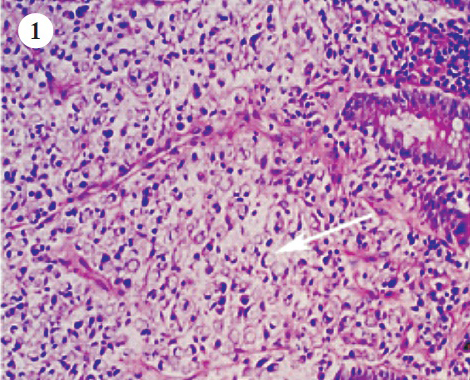
- Histopathological examination revealed the colonic signet-ring cell carcinoma (arrow) (Hematoxylin-eosin staining; original magnification×200).
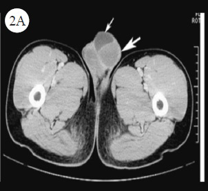
- CT showed the only hydrocele of tunica vaginalis (thin arrow) and the normal shape of testis (thick arrow) in the left scrotum during December 2010.
- CT showed a 6.8×4.5cm mixed density mass shadow with ill-defined margin in the left scrotum, and above which a cystic shadow with watery density in May 2014. CT diagnosis was left testis tumour (thick arrow) and hydrocele of tunica vaginalis (thin arrow).
-
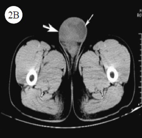
A-D. Immunohistochemistry (IHC) staining showed that the tumour cells were positive for cytokeratin (CK) 20 (A) (arrow), Villin (B) (arrow), caudal-related homeobox transcription factor 2 (CDX2) (C) (arrow), but negative for CK7 (D) (arrow) (DAB staining; original magnification ×200).
-
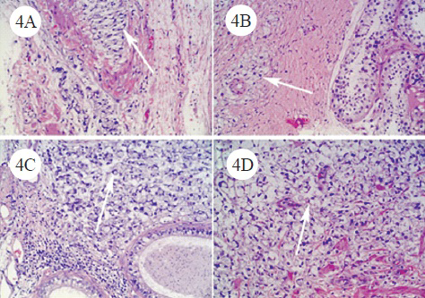
A, B, C, D. Histopathological examination revealed metastatic colonic signet-ring cell carcinoma to the left tunica vaginalis testis (A) (arrow), tunica albuginea testis (B) (arrow), epididymis (C) (arrow) and spermatic cord (D) (arrow) (Hematoxylin-eosin staining; original magnification×200).
Acknowledgment
Authors thank chief physician Rui-zhen Hong and attending physician Li-wen Xu from the Deparment of Radiology, The Dingli Clinical Institute of Wenzhou Medical University (Wenzhou Central Hospital), Wenzhou, Zhejiang, PR China, for assistance in the evaluation of CT.


