Translate this page into:
Arc of Riolan
* For correspondence: binitsurekapgi@gmail.com
This is an open-access article distributed under the terms of the Creative Commons Attribution-Noncommercial-Share Alike 3.0 Unported, which permits unrestricted use, distribution, and reproduction in any medium, provided the original work is properly cited.
This article was originally published by Medknow Publications & Media Pvt Ltd and was migrated to Scientific Scholar after the change of Publisher.
A 55-yr old woman presented to the Department of Medicine at Vardhman Mahavir Medical College & Safdarjung Hospital, New Delhi, India, in May, 2012 with symptoms of abdominal angina, and 5 kg weight loss over the last eight weeks. She had no history of jaundice or melena. On examination, abdomen was soft with no organomegaly. Laboratory workup showed anaemia with haemoglobin 8.4 g/dl, and normal white cell and platelet count. Contrast-enhanced CT abdominal angiography revealed severe occlusive atherosclerotic aortoiliac disease, non-visualization of infrarenal abdominal aorta with Arc of Riolan (AOR) connecting superior mesenteric and inferior mesenteric arteries (SMA and IMA) (Figs. 1, 2). She was put on antispasmodics and was advised for endovascular surgery. However, the patient has refused surgery and is on follow up.
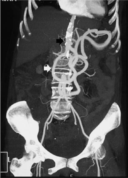
- CT abdominal angiography maximum-intensity projection (MIP) image showing atherosclerotic occlusive aortoiliac disease (black arrow) with Arc of Riolan (white arrow) connecting SMA and IMA.
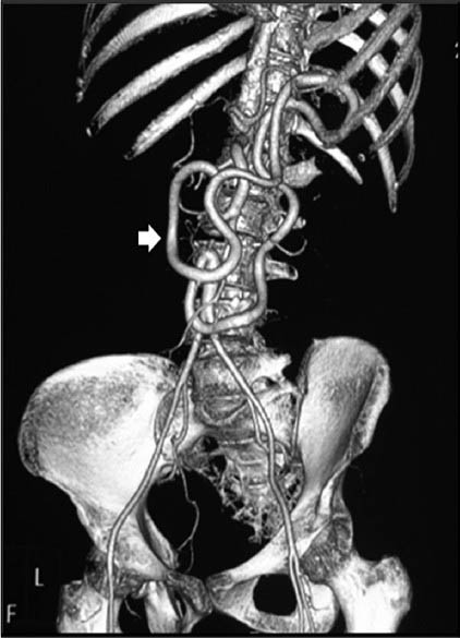
- Volume-rendered 3D CT angiography image better depicting large mesenteric collateral Arc of Riolan (arrow).
AOR, named after Jean Riolan, a French anatomist 1, is also known as meandering mesenteric/central anastomotic artery. It connects middle colic branch of SMA with left colic branch of IMA that runs close to the root of the mesentery 2. In SMA/IMA/distal aortic occlusion, the AOR provides collateral flow between IMA and SMA 23. CT angiography is the investigation of choice in such cases as it can visualize surrounding organs, minimally invasive, allows 3D image reformatting and has a higher sensitivity and specificity 2. Another such collateral is the marginal artery of Drummond. Identification of this rare anatomic variation is important for intervention radiologists.
References
- The variant blood supply to the descending colon, rectosigmoid and rectum based on 400 dissections. Its importance in regional resections: a review of medical literature. Dis Colon Rectum. 1965;8:251-78.
- [Google Scholar]
- Volume-rendered 3D CT of the mesenteric vasculature: normal anatomy, anatomic variants, and pathologic conditions1. Radiographics. 2002;22:161-72.
- [Google Scholar]
- Role of preoperative angiography in colon interposition surgery. Diagn Interv Radiol. 2012;18:314-8.
- [Google Scholar]


