
Translate this page into:
An uncommon presentation of strongyloidiasis
*For correspondence: medsuhas1987@gmail.com
-
Received: ,
This is an open access journal, and articles are distributed under the terms of the Creative Commons Attribution-NonCommercial-ShareAlike 4.0 License, which allows others to remix, tweak, and build upon the work non-commercially, as long as appropriate credit is given and the new creations are licensed under the identical terms.
This article was originally published by Wolters Kluwer - Medknow and was migrated to Scientific Scholar after the change of Publisher.
A 30 yr old immunocompetent male† presented with anasarca of one month to the department of Gastroenterology, Topiwala National Medical College & B.Y.L. Nair Charitable Hospital, Mumbai, India, in March 2019. On evaluation, haemoglobin (10.9 g/dl) and serum protein (2.8 g/dl) were low; other laboratory findings were normal. Computed tomographic scan of the abdomen showed jejunoileal fold pattern reversal with mesenteric lymphadenopathy (Fig. 1); upper gastrointestinal endoscopy showed duodenal fissuring (Fig. 2A); laparoscopy showed prominent mesenteric lymph nodes (Fig. 2B); histopathological examination showed larvae and eggs of Strongyloides (Fig. 3A-C). A diagnosis of strongyloidiasis complicating protein-losing enteropathy was made.Faecal alpha 1-antitrypsin clearance suggested protein-losing enteropathy. The patient was treated with two days of oral ivermectin and was asymptomatic on seven months of follow up.
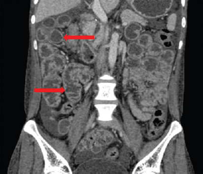
- Contrast-enhanced computed tomography scan of abdomen showing jejunoileal folds pattern reversal: Featureless jejunal loops (upper red arrow) and increased mucosal folds in the ileal loops (lower red arrow).
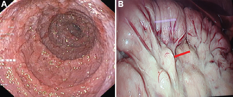
- (A) Upper gastrointestinal endoscopic picture showing severe duodenal fissuring. (B) Laparoscopic picture showing ileal loops with enlarged mesenteric lymph nodes (red arrow).
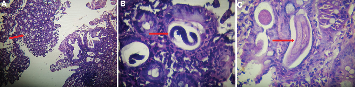
- (A) Histopathology photomicrograph showing duodenal mucosa with multiple eggs of Strongyloides stercoralis (H and E, ×10). (B) Histopathology photomicrograph showing filariform larvae of Strongyloides stercoralis (H and E, ×40). (C) Histopathology photomicrograph showing rhabditiform larvae of Strongyloides stercoralis (H and E, ×40).
Acknowledgment:
Authors thank Dr Rima Kamat, department of Pathology, Topiwala National Medical College & B.Y.L. Nair Charitable Hospital, Mumbai, for making histopathologic diagnosis and providing histopathology images for the case presented.
Conflicts of Interest: None.


