
Translate this page into:
Acute abdomen in a patient with chronic myeloid leukaemia
*For correspondence: drshraddhapatkar@gmail.com
-
Received: ,
This is an open access journal, and articles are distributed under the terms of the Creative Commons Attribution-NonCommercial-ShareAlike 4.0 License, which allows others to remix, tweak, and build upon the work non-commercially, as long as appropriate credit is given and the new creations are licensed under the identical terms.
This article was originally published by Wolters Kluwer - Medknow and was migrated to Scientific Scholar after the change of Publisher.
A 29 yr old male†, diagnosed with chronic myeloid leukaemia (CML), presented to the department of Surgical Oncology, Tata Memorial Hospital, Mumbai, India, in September 2019, with acute abdominal pain and distension. He had tachycardia with diffuse abdominal tenderness and guarding. Haemoglobin value was 4.8 g/dL with total leukocyte count of 124,000/mm3. Computed tomography scan revealed a large peritoneal collection with areas of haemorrhage, suspected arterial feeder from the anterior abdominal wall (Fig. 1A), with scalloping of the left lobe of the liver (Fig. 1B). He underwent angiography, which showed no active bleeding source. The patient later underwent exploratory laparotomy, which revealed a ruptured abscess in the left lobe of the liver and a left lateral sectionectomy was performed (Fig. 2). Histopathology showed an abscess with filamentous bacteria negative for special stains (Fig. 3). At one month of follow up, patient was clinically doing well.
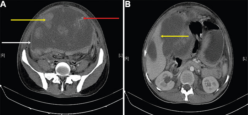
- (A) Contrast-enhanced computed tomography abdomen showing large heterogeneous cystic lesion (white arrow) with multiple solid appearing (yellow arrow), irregular, non-enhancing densities within. Also noted is a vessel arising from the anterior abdominal wall (red arrow) (suspected source of haemoperitoneum). (B) Contrast-enhanced computed tomography abdomen showing the cystic lesion scalloping the left lobe of liver (arrow).

- Gross specimen of the left lateral sectionectomy showing a ruptured liver abscess (arrow).
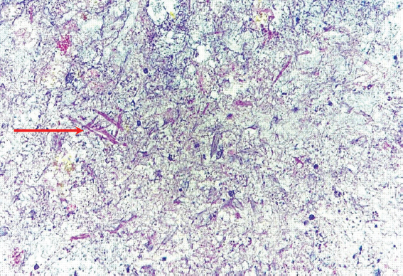
- High-power magnification (×400) of H and E-stained section showing filamentous bacteria (arrow) in a background of necrosis and inflammation. The organisms were negative for special stains.
Conflicts of Interest: None.


