
Translate this page into:
Abject health concern in elderly: Cutaneous metastasis of malignant melanoma of anal canal
*For correspondence: drneelam428@yahoo.co.in
-
Received: ,
This is an open access journal, and articles are distributed under the terms of the Creative Commons Attribution-NonCommercial-ShareAlike 4.0 License, which allows others to remix, tweak, and build upon the work non-commercially, as long as appropriate credit is given and the new creations are licensed under the identical terms.
This article was originally published by Wolters Kluwer - Medknow and was migrated to Scientific Scholar after the change of Publisher.
A 76 yr old emaciated male† presented to department of Surgery, University College of Medical Sciences and Guru Teg Bahadur Hospital, Delhi, India, in December 2016, with multiple distinctly bluish to blackish, non-pulsatile, firm-to-hard nodules ranging in size from 0.4 to 1.2 cm over anterior abdominal wall and back and left inguinal lymphadenopathy for past six months (Fig. 1A and B). Angiosarcoma of the skin and cutaneous metastasis was suspected clinically. Fine-needle aspiration cytology of cutaneous lesions and lymph node demonstrated highly pigmented pleomorphic cells suspicious of malignant melanoma (Fig. 2A and B). On probing the patient embarrassingly admitted to having large foul-smelling anal ulcerated mass lesion (Fig. 1C) for the past three years. Histologic findings of the biopsy of cutaneous lesions and anal mass showed strong immunopositivity for human melanoma black (HMB)-45, confirming diagnosis of malignant melanoma with cutaneous metastasis (Fig. 2C and D and inset). The advanced tumour stage precluded surgical intervention, so the family opted for no treatment in view of grim prognosis.
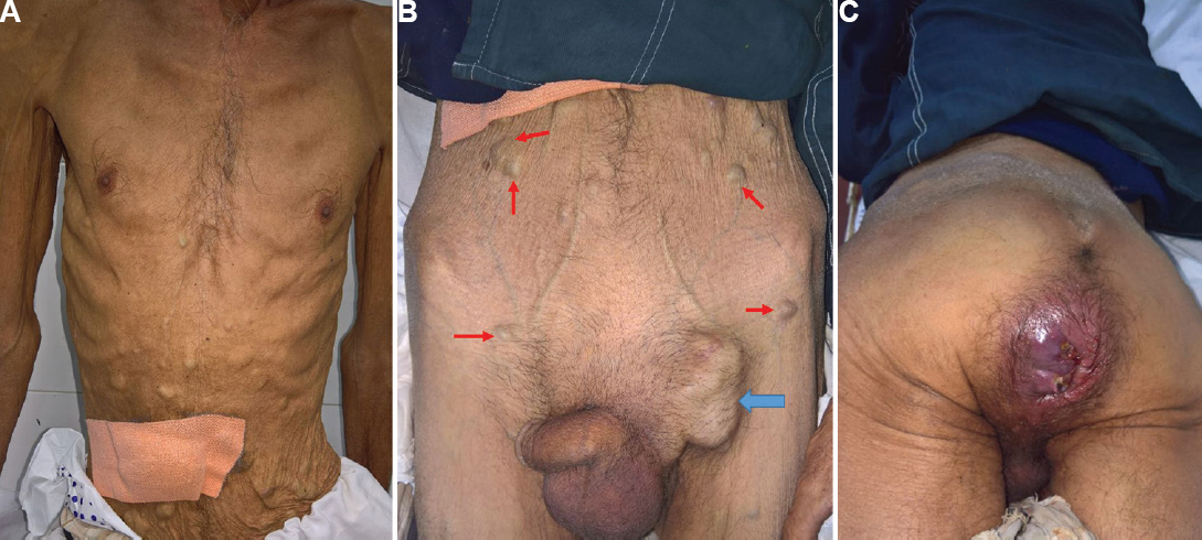
- (A) Multiple bluish cutaneous nodules over anterior abdominal wall (red arrows). (B) enlarged left inguinal lymph nodes (blue arrows) and (C) ulcerated anal mass.
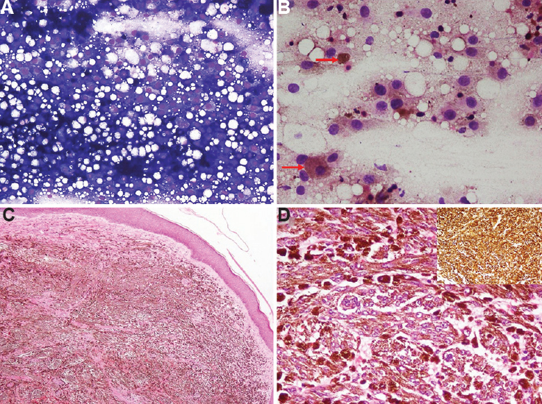
- (A and B) FNA smears from cutaneous nodules showing sheets of pleomorphic cells with intracytoplasmic melanin pigment (red arrows) (May Grunwald Giemsa, ×200 and ×400 respectively). (C) Histopathology of skin nodule showing metastatic malignant melanoma with flattening of epidermis (H and E, ×100) and (D) Histopathology of anal lesion (H and E, ×400), (inset) Immunostain for HMB-45 (×400).
Acknowledgment:
Authors acknowledge Dr Sanjay Gupta, department of Surgery, UCMS & GTB Hospital, Delhi, for referring the patient.
Conflicts of Interest: None.


