Translate this page into:
A rare case of leiomyomatosis peritonealis disseminata masquerading as peritoneal carcinomatosis
*For correspondence: doc_rj24@yahoo.com
-
Received: ,
This is an open access journal, and articles are distributed under the terms of the Creative Commons Attribution-NonCommercial-ShareAlike 4.0 License, which allows others to remix, tweak, and build upon the work non-commercially, as long as appropriate credit is given and the new creations are licensed under the identical terms.
This article was originally published by Wolters Kluwer - Medknow and was migrated to Scientific Scholar after the change of Publisher.
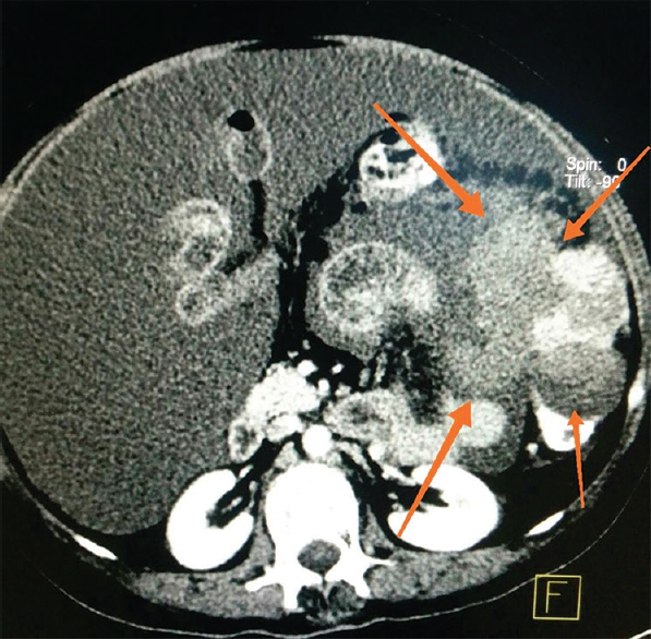
A 58 yr old postmenopausal woman† presented to the department of Surgical Oncology, Dayanand Medical College & Hospital, Ludhiana, India, in July 2016, with lower abdominal pain and an antecedent history of total abdominal hysterectomy with bilateral salpingo-oophorectomy done for uterine leiomyoma and ovarian fibroma. Although radiology was suggestive of disseminated intraperitoneal malignancy (Fig. 1), histopathological (Fig. 2A) and immunohistochemical examination showed positivity for smooth muscle actin (Fig. 2B) and oestrogen (Fig. 2C) and progesterone (Fig. 2D) receptors, thus revealing leiomyomatosis peritonealis disseminata which grossly mimiced disseminated malignancy but was histologically benign otherwise. Plan for surgery was deferred, and the patient was put on tamoxifen. At one year follow up, the patient remained asymptomatic without any radiographic disease progression. A pathological evaluation is crucial for the diagnosis, surgery is not always needed and close surveillance is a necessity rather than a recommendation.

- Contrast-enhanced computed tomography axial section of the abdomen showing a heterogeneous enhancing soft-tissue lesion (arrows) in the left lumbar region along the serosal surface of the left colon with necrotic areas within it.

- (A) Benign mesenchymal tumour composed of oval-to-spindle-shaped cells having bland nuclear features and scanty cytoplasm (H and E, ×40); immunohistochemistry in neoplastic cells showing membranous positivity for (B) smooth muscle actin (arrow; x40); (C) nuclear positivity for oestrogen receptor (arrow; x40) and (D) progesterone receptor (arrow; x40).
Conflicts of Interest: None.


