
Translate this page into:
A rare case of cerebrotendinous xanthomatosis with typical clinical & radiological features
*For correspondence: sanghamitralaskar@rediffmail.com
-
Received: ,
This article was originally published by Wolters Kluwer - Medknow and was migrated to Scientific Scholar after the change of Publisher.
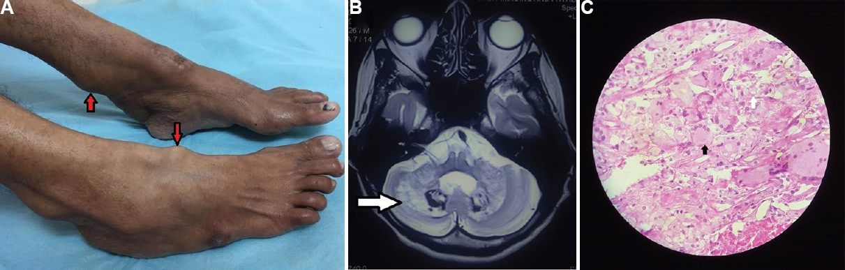
A 35 yr old woman† presented to the department of Neurology, Safdarjung Hospital, New Delhi, in May 2019, with a history of bilateral cataract surgery at the age of 17 yr followed by declining academic performance starting at the same age, cutaneous swellings since the age of 25 yr along with gait ataxia, recurrent falls, difficulty in opening her mouth and frequent crying spells since the age of 33 yr. She had multiple painless firm globular swellings without overlying skin changes over bilateral ankles and feet (Figure A). Neurological examination revealed executive dysfunction, perseveration and visuospatial dysfunction with slow saccades, broken pursuit, reduced palatal movements, slurred speech, limb and gait ataxia and jaw-closing dystonia. Magnetic resonance imaging brain revealed high-signal intensity areas on T2-weighted and FLAIR images in the bilateral dentate nuclei and cerebellar white matter, suggestive of accumulation of sterols in the nerve cells along with varying degrees of demyelination (Figure B). Histopathology of the cutaneous swellings revealed foamy histiocytes admixed with giant cells with multiple nuclei arranged in a garland-like fashion, along with a large number of extracellular cholesterol crystals in the background suggestive of tendon xanthomas (Figure C). Based on the typical features and histopathology, she was diagnosed with cerebrotendinous xanthomatosis. She was treated with ursodeoxycholic acid and botulinum toxin injection into the masseter and showed improvement in jaw dystonia at 10 months follow up.
-
(A) Firm globular tendon xanthomas without overlying skin changes over both ankles and feet (arrows), (B) T2-weighted magnetic resonance imaging showing high-signal intensity areas in the bilateral dentate nuclei and cerebellar white matter (arrow), (C) histopathological examination of tendon xanthoma showing foamy histiocytes (arrow) admixed with giant cells (black arrow) having multiple nuclei arranged in a garland-like fashion, with a large number of extracellular cholesterol crystals in the background conclusive of tendon xanthomas (H and E stain, ×100).
Conflicts of Interest: None.


