
Translate this page into:
A cap over the kneecap
*For correspondence: drganeshortho@gmail.com
-
Received: ,
This is an open access journal, and articles are distributed under the terms of the Creative Commons Attribution-NonCommercial-ShareAlike 4.0 License, which allows others to remix, tweak, and build upon the work non-commercially, as long as appropriate credit is given and the new creations are licensed under the identical terms.
This article was originally published by Wolters Kluwer - Medknow and was migrated to Scientific Scholar after the change of Publisher.
A 65 yr old female† presented to the department of Orthopaedics, Government Medical College, Haldwani, Uttarakhand, India, in January 2018, with an atraumatic, old hard bony lump over the right knee without pain, functional limitation or other 'red flags' (Fig. 1A and B). Radiography showed a radio-opaque, sclerotic mass over the patella (Fig. 1C). Magnetic resonance imaging showed a mass with calcified matrix overlying the patella like a tilted bony cap without any patellar affliction (Fig. 2). The mass was surgically excised. The excision biopsy of the mass yielded a bony hard mass with variegated surface (Fig. 3A and B), however, the patella was unaffected (Fig. 3C). Histological diagnosis was extra-skeletal osteochondroma (Fig. 3D). No complication and recurrence were noted in the follow up of nine months. Knee is not a preferred site for these lesions and most present as smaller growths. A giant extra-skeletal osteochondroma is rarity, and its dormant existence over a bone like an outer envelope is a striking presentation.
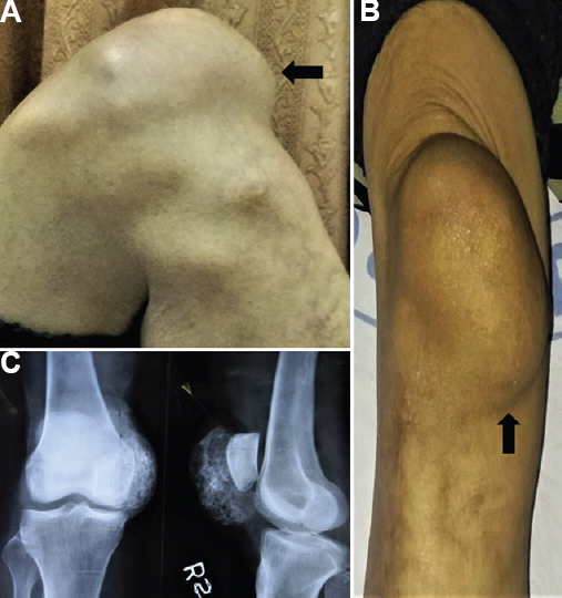
- (A and B) The clinical image of a lump over the patella with no effect on knee flexion and extension (arrows). (C) The radiograph showing a lesion with well-defined radio-opacity overlying patella which appears to have neither apparent association with patella nor any anatomical alteration in the patella noted.
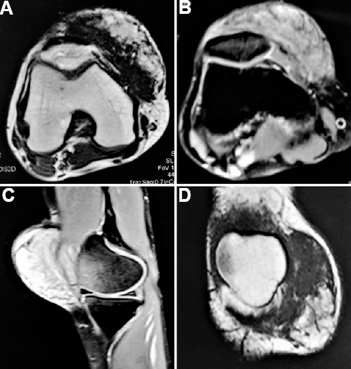
- The axial (A) T1-weighted and (B) T2-weighted magnetic resonance images showing the presence of mass overlying patella, more on the medial aspect, with calcified matrix and intact patellar anatomy. (C) Sagital images on the medial aspect of the knee showing the mass and (D) coronal magnetic resonance images delineating the lesion as a mass with heterogeneous calcified matrix.
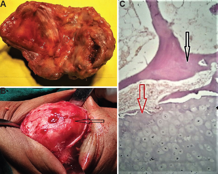
- (A) The inner aspects of the excised mass (about 8 cm × 6 cm) with variegated appearance and bony hard. (B) The intact patella (arrow), following the excision of the mass, with no remnants of the same denoting that the lesion existed as a separate overlying structure. (C) The histological features showing the presence of bony elements (black arrow) (H and E, ×10) and varying thickness of cartilage cap layer (red arrow) over its outer surface with bluish tinge, suggestive of an extra-skeletal osteochondroma.
Conflicts of Interest: None.


