
Translate this page into:
Flowers in the sky & lumps on the ground
*For correspondence: dr_vimalkpaliwal@rediffmail.com
-
Received: ,
This is an open access journal, and articles are distributed under the terms of the Creative Commons Attribution-NonCommercial-ShareAlike 4.0 License, which allows others to remix, tweak, and build upon the work non-commercially, as long as appropriate credit is given and the new creations are licensed under the identical terms.
This article was originally published by Wolters Kluwer - Medknow and was migrated to Scientific Scholar after the change of Publisher.
A 36 yr old male† was presented to the department of Neurology, Sanjay Gandhi Postgraduate Institute of Medical Sciences, Lucknow, India, in August 2019, with acute daily headache and irritable behaviour for one month. He suffered from inadequately treated epilepsy for 20 years. On examination, multiple small subcutaneous nodules were observed all over his body. His neurological examination was normal. His cranial magnetic resonance imaging (MRI) showed multiple large bilateral parenchymal cystic racemose neurocysticerci (Figure A-C). The ultrasound of subcutaneous nodules showed cysticercus with a scolex (Figure D). The patient was treated with albendazole for four weeks along with oral prednisolone. The patient's symptoms improved promptly with steroids as he was asymptomatic at three months follow up.
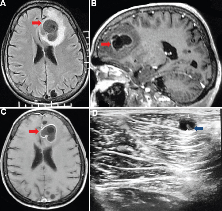
- (A) Cranial magnetic resonance imaging (fluid attenuated inversion recovery sequence) showing a large left frontal racemose neurocysticercal lesion (red arrow) with eccentric calcification and peri-focal oedema. (B and C) Sagittal and axial post-contrast T1-weighted images showing ring-enhancing racemose neurocysticercal lesion (red arrow) with few other small ring-enhancing cystic lesions in the left frontal and occipital lobe. (D) Ultrasound of the left thigh lesion showing a cystic lesion with central scolex (blue arrow).
Racemose neurocysticerci are large confluent cystic masses usually seen in the subarachnoid or intraventricular spaces. Multiple neurocysticerci are readily diagnosed on MRI. Being rare, large parenchymal racemose neurocysticerci may be confused with other helminthic cysts or cystic neoplasms.
Conflicts of Interest: None.


