
Translate this page into:
Masquerading verrucous carcinoma: A pathologist's & surgeon's dilemma
*For correspondence: dr_shaluk@rediffmail.com
-
Received: ,
This is an open access journal, and articles are distributed under the terms of the Creative Commons Attribution-NonCommercial-ShareAlike 4.0 License, which allows others to remix, tweak, and build upon the work non-commercially, as long as appropriate credit is given and the new creations are licensed under the identical terms.
This article was originally published by Wolters Kluwer - Medknow and was migrated to Scientific Scholar after the change of Publisher.
An 80 yr old male† presented to the department of Oral & Maxillofacial Surgery, AB Shetty Memorial Institute of Dental Sciences, Mangaluru, Karnataka, India, in March 2018, with a slow-growing (two years), single, whitish-pink, well-defined, exophytic growth on the left buccal mucosa approximately 3 × 3 cm in size (Fig. 1). On palpation, the lesion was tender, firm in consistency, with irregular surface, no fixity and no palpable lymph nodes. A provisional diagnosis of verrucous carcinoma was made which was confirmed on incisional biopsy (Fig. 2), and the patient was taken up for wide excision and reconstruction with local flap. However, to our surprise, histopathology of this innocuous lesion revealed features of papillary squamous cell carcinoma (Fig. 3). The patient underwent radiotherapy and was on regular follow up; however, in March 2019, he reported with recurrence. Verrucous carcinoma can pose a diagnostic dilemma for both surgeon and pathologist. Histopathologic grading and interdisciplinary discussions may help in preventing over- or under-treatment.
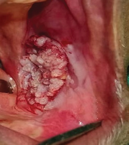
- Clinical image showing single, exophytic cauliflower-like whitish-pink-coloured, well-defined growth on the left buccal mucosa approximately measuring 3 × 3 cm in size extending to the buccal vestibule.
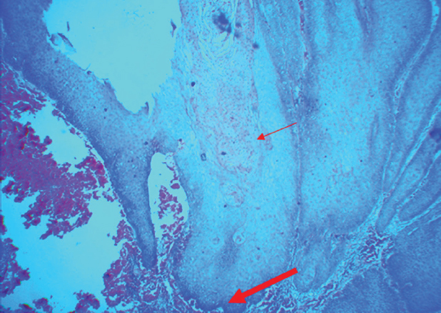
- Photomicrograph of the incisional biopsy showing stratified squamous epithelium with papillary masses, keratin plugging (thin red arrow) and blunt and broad rete pegs with underlying chronic inflammatory cells (thick red arrow) in the connective tissue (H and E, ×4).
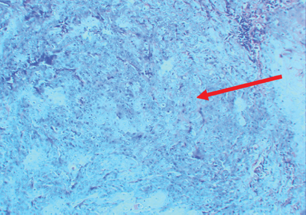
- Photomicrograph of the excisional biopsy showing dysplastic epithelium proliferating into the connective tissue (red arrow) suggestive of papillary squamous cell carcinoma (H and E, ×40).
Conflicts of Interest: None.


