
Translate this page into:
A rare case of bilateral calcified testicular mass
*For correspondence: slims.surgery@gmail.com
-
Received: ,
This is an open access journal, and articles are distributed under the terms of the Creative Commons Attribution-NonCommercial-ShareAlike 4.0 License, which allows others to remix, tweak, and build upon the work non-commercially, as long as appropriate credit is given and the new creations are licensed under the identical terms.
This article was originally published by Wolters Kluwer - Medknow and was migrated to Scientific Scholar after the change of Publisher.
A 78 yr old male† patient came to the department of General Surgery, Sri Lakshmi Narayana Institute of Medical Sciences, Puducherry, India, in August 2019, with complaints scrotal swelling of the right side for the past five years with initial size of 3 × 3 cm which gradually increased to 8 × 6 cm. The swelling was hard in consistency and the testis was not palpable skin rugosity was also present. He also noticed left-sided scrotal swelling for two years. No history suggestive of malignancy or trauma was noted. He had undergone left-sided hydrocele surgery 15 yr back. He was not an alcoholic or a smoker. Left scrotal swelling was 5 × 3 cm in size, with similar features. No palpable iliac nodes were noted. He was diagnosed with a bilateral testicular mass suspicious of malignancy. X-ray showed enlarged right testis with bilateral testicular peripheral rim calcification (Fig. 1). Computed tomographic scan showed no para-aortic lymphadenopathy (Fig. 2). Bilateral orchiectomy was done (Fig. 3). Histopathological examination was suggestive of pyocele with calcification.
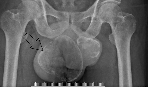
- Plain X-ray pelvis showing peripheral rim of calcification of bilateral testis (arrow).
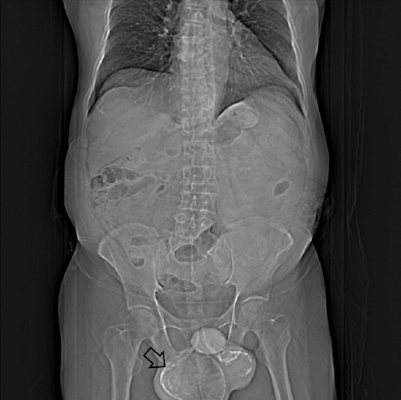
- Plain computed tomographic abdomen and pelvis showing peripheral rim of calcification of bilateral testis with no para-aortic lymphadenopathy.
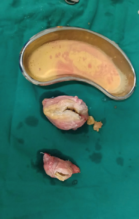
- Postoperative specimen showing both testes with pyocele.
Conflicts of Interest: None.


